
Pilonidal disease
| Pilonidal disease | |
|---|---|
| Other names | Pilonidal cyst, pilonidal abscess, pilonidal sinus, sacrococcygeal cyst / fistula |
surgical removal | |
| Frequency | 3 per 10,000 per year[2] |
Pilonidal disease is a type of skin infection which typically occurs as a cyst between the cheeks of the buttocks and often at the upper end.[1][3] Symptoms may include pain, swelling, and redness.[1] There may also be drainage of fluid, but rarely a fever.[1][2]
Risk factors include
If there is infection, treatment is generally by
About 3 per 10,000 people per year are affected, and it occurs more often in males than females.[2] Young adults are most commonly affected.[2] The term pilonidal means 'nest of hair'.[1] The condition was first described in 1833.[1]
Signs and symptoms

Pilonidal cysts can be itchy and often very painful, and typically occur between the ages of 15 and 35.[5] Although usually found near the coccyx, the condition can also affect the navel, armpit, the cheek,[6] or the genital region,[7] though these locations are much rarer.
Signs and symptoms may include:[8]
- Intermittent pain/discomfort or swelling above the anus or near the tailbone
- Opaque yellow (purulent) or bloody discharge from the tailbone area
- Unexpected moisture in the tailbone region
- Discomfort sitting on the tailbone, doing sit-ups or riding a bicycle—any activities that roll over the tailbone area
Some people with a pilonidal cyst will be asymptomatic.[9]
Pilonidal sinus
Pilonidal sinus (PNS): is a sinus tract, or small channel, that may originate from the source of infection and open to the surface of the skin.[10] Material from the cyst drains through the pilonidal sinus. A pilonidal cyst is usually painful, but if it is a draining sinus, the pressure is relieved and patient might not feel pain.
Causes
Hair insertion is the causative agent of pilonidal cysts.[11][12] An analysis of 624 patients' cyst hair found that 74% of the hair was rootless, and resembled spiky, razor-cut hair rather than intact body hair.[11] One proposed cause is ingrown hair,[13] although hairs found in pilonidal sinus tracts have more often been found to originate from the head.
Excessive sitting is thought to predispose people to the condition, as sitting increases pressure on the
Trauma is not believed to cause a pilonidal cyst; however, such an event may result in inflammation of an existing cyst; there are cases where this can occur months after a localized injury to the area.
Pilonidal cysts may be caused by a congenital pilonidal dimple.[14]
Excessive sweating can also contribute to the formation of a pilonidal cyst: moisture can fill a stretched hair follicle, which helps create a low-oxygen environment that promotes the growth of
Differential diagnosis

A pilonidal cyst can resemble a
Treatment
If there is infection, treatment is generally by
The evidence for elective treatment of pilonidal sinus disease is poor.[17] The most commonly performed surgery is for the pilonidal sinus complex to be surgically excised with the wound often left open to heal. Post-surgical wound packing may be necessary, and packing typically must be replaced daily for four to eight weeks. In some cases, two years may be required for complete granulation to occur. Sometimes the cyst is resolved via surgical marsupialization.[18]
A 2018 literature review of 740 records of surgeries that included recurrence rates found that primary midline closure surgeries resulted in a 67.9% recurrence rate within 20 years, and recommended that they should be discontinued due to the high recurrence rate.[19] Incision and drainage had a recurrence rate of 25.9% within 2 years, up to 40.2% in 5 years. Phenol treatment has a recurrence rate of 14.1% at 2 years and 40.4% at 5 years.[19]
Surgeons can also excise the sinus and repair with a reconstructive flap technique, such as a "cleft lift" procedure or Z-plasty, usually done under
Another technique is to treat pilonidal sinus with fibrin glue. This technique is of unclear benefit as of 2017 due to insufficient research.[22] The evidence for any treatment is of low quality, and care must be taken not to over interpret any study in this field.[17]
Since the 2010s, a number of minimally invasive techniques have been developed, with the purpose of minimising the impact of surgery on patients and of achieving less pain and shorter recovery times.[23]
In some cases, the wounds are left open after surgery to heal naturally instead of being closed with stitches. There are a lot of different dressings and topical agents (creams or lotions) that are available for helping these open wounds to heal. A 2022 systematic review brought together evidence from 11 studies that compared dressings and topical agents for treating open wounds after surgical treatment for pilonidal sinus of the buttocks.[24] The authors concluded that: platelet rich plasma may help wounds to heal quicker compared to sterile gauze; Lietofix skin repair cream may help wounds to heal by 30 days compared to iodine (which helps to reduce bacteria in the wound); but it is not clear whether hydrogel dressings (designed to keep the wound moist) reduce the time it takes wounds to heal compared with cleaning the wound with iodine.[24]
Endoscopic pilonidal treatment, which uses a small camera to guide the surgeon in removing hair, is a newer method of treatment that has minimal pain and quick healing compared to surgery. A literature review of 497 patients found that the average endoscopic operation time was 34.7 minutes, and the average healing time was 32.9 days. Failure occurred in 8% of patients, who had persistent disease or recurrence.[25]
-
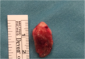 Excised pilonidal cyst
 -
 Trephine/biopsy punch minimally invasive surgery for pilonidal disease (1)
 -
 Trephine/biopsy punch minimally invasive surgery for pilonidal disease (2)
 -
 Pilonidal cyst two days after traditional closed surgery.
 -
 Anatomy of pilonidal disease removed after trephine or biopsy punch surgery: pilonidal fistula (top) and pilonidal cyst (bottom)


Etymology
Pilonidal means 'nest of hair' and is derived from the Latin words for 'hair' (pilus) and 'nest' (nidus).[5] The condition was first described by Herbert Mayo in 1833.[26] R. M. Hodges was the first to use the phrase pilonidal cyst to describe the condition in 1880.[27][28]
The condition was widespread in the United States Army during World War II. The condition was termed "Jeep seat" or "Jeep riders' disease", because a large portion of people who were being hospitalized for it rode in Jeeps, and prolonged bumpy rides in the vehicles were believed to have caused the condition due to irritation and pressure on the coccyx.
References
- ^ PMID 22379405.
- ^ ISBN 9780323529570. Archivedfrom the original on 2023-01-14. Retrieved 2020-09-20.
- ISBN 9780323319690. Archivedfrom the original on 2023-01-14. Retrieved 2020-09-20.
- ^ "Pilonidal Cyst Laser Hair Removal". Cure Pilonidal Cyst. Archived from the original on 2022-07-06. Retrieved 2021-08-23.
- ^ a b "Pilonidal Cyst: Definition". Mayo Clinic. December 5, 2012. Archived from the original on December 27, 2012. Retrieved February 8, 2013.
- PMID 33563340.
- S2CID 30090441.
- ^ Sternberg J. "What Is Pilonidal Disease". Archived from the original on November 11, 2014. Retrieved November 14, 2014.
- ^ Doerr S. "Pilonidal Cyst". eMedicineHealth. p. 1. Archived from the original on February 12, 2013. Retrieved February 8, 2013.
- ^ "PNS Acronyms". thefreedictionary.com. Archived from the original on 2018-01-04. Retrieved 2018-01-03.
- ^ S2CID 4705771.
- PMID 1575660.
- ^ "Pilonidal Cyst: Causes". Mayo Clinic. December 5, 2012. Archived from the original on October 25, 2008. Retrieved February 8, 2013.
- S2CID 189796851.
- ^ PMID 12361421.
- PMID 24913119.
- ^ PMID 31754976.
- S2CID 206172705.
- ^ PMID 29449548.
- PMID 20091589.
- S2CID 88464026.
- PMID 28085995.
- PMID 35344244.
- ^ PMID 35593897.
- S2CID 4472852.
- ^ Lanigan M (September 27, 2012). "Pilonidal Cyst and Sinus". Medscape. WebMD. Archived from the original on October 22, 2008. Retrieved February 8, 2013.
- (PDF) from the original on 2021-08-28. Retrieved 2020-02-10.
- ISBN 3-540-64046-0.