Tracheal intubation
| Tracheal intubation | |
|---|---|
airway anatomy | |
| ICD-9-CM | 96.04 |
| MeSH | D007442 |
| OPS-301 code | 8-701 |
| MedlinePlus | 003449 |
Tracheal intubation, usually simply referred to as
The most widely used route is orotracheal, in which an endotracheal tube is passed through the mouth and vocal apparatus into the trachea. In a nasotracheal procedure, an endotracheal tube is passed through the nose and vocal apparatus into the trachea. Other methods of intubation involve surgery and include the cricothyrotomy (used almost exclusively in emergency circumstances) and the tracheotomy, used primarily in situations where a prolonged need for airway support is anticipated.
Because it is an
After the trachea has been intubated, a balloon cuff is typically inflated just above the far end of the tube to help secure it in place, to prevent leakage of respiratory gases, and to protect the
For centuries,
Tracheal intubation can be associated with
Indications
Tracheal intubation is
Depressed level of consciousness
Perhaps the most common indication for tracheal intubation is for the placement of a conduit through which
Damage to the brain (such as from a massive
Hypoxemia
Intubation may be necessary for a patient with
Airway obstruction
Actual or impending airway obstruction is a common indication for intubation of the trachea. Life-threatening airway obstruction may occur when a
Manipulation of the airway
Diagnostic or therapeutic manipulation of the airway (such as bronchoscopy, laser therapy or stenting of the bronchi) may intermittently interfere with the ability to breathe; intubation may be necessary in such situations.[4]
Newborns
Syndromes such as
Equipment
Laryngoscopes


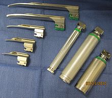
The vast majority of tracheal intubations involve the use of a
The decision to use a straight or curved laryngoscope blade depends partly on the specific anatomical features of the airway, and partly on the personal experience and preference of the laryngoscopist. The Miller blade, characterized by its straight, elongated shape with a curved tip, is frequently employed in patients with challenging airway anatomy, such as those with limited mouth opening or a high larynx. Its design allows for direct visualization of the epiglottis, facilitating precise glottic exposure.[10] Conversely, the Macintosh blade, with its curved configuration reminiscent of the letters "C" or "J," is favored in routine intubations for patients with normal airway anatomy. Its curved design enables indirect laryngoscopy, providing enhanced visualization of the vocal cords and glottis in most adult patients.[11]
The choice between the Miller and Macintosh blades is influenced by specific anatomical considerations and the preferences of the laryngoscopist. While the Macintosh blade is the most commonly utilized curved laryngoscope blade, the Miller blade is the preferred option for straight blade intubation. Both blades are available in various sizes, ranging from size 0 (infant) to size 4 (large adult), catering to patients of different ages and anatomies. Additionally, there exists a myriad of specialty blades with unique features, including mirrors for enhanced visualization and ports for oxygen administration, primarily utilized by anesthetists and otolaryngologists in operating room settings.[12][10]
Stylets

An intubating stylet is a malleable metal wire designed to be inserted into the endotracheal tube to make the tube conform better to the upper airway anatomy of the specific individual. This aid is commonly used with a difficult laryngoscopy. Just as with laryngoscope blades, there are also several types of available stylets,[17] such as the Verathon Stylet, which is specifically designed to follow the 60° blade angle of the GlideScope video laryngoscope.[18]
The Eschmann tracheal tube introducer (also referred to as a "gum elastic bougie") is specialized type of stylet used to facilitate difficult intubation.[19] This flexible device is 60 cm (24 in) in length, 15 French (5 mm diameter) with a small "hockey-stick" angle at the far end. Unlike a traditional intubating stylet, the Eschmann tracheal tube introducer is typically inserted directly into the trachea and then used as a guide over which the endotracheal tube can be passed (in a manner analogous to the Seldinger technique). As the Eschmann tracheal tube introducer is considerably less rigid than a conventional stylet, this technique is considered to be a relatively atraumatic means of tracheal intubation.[20][21]
The tracheal tube exchanger is a hollow catheter, 56 to 81 cm (22.0 to 31.9 in) in length, that can be used for removal and replacement of tracheal tubes without the need for laryngoscopy.[22] The Cook Airway Exchange Catheter (CAEC) is another example of this type of catheter; this device has a central lumen (hollow channel) through which oxygen can be administered.[23] Airway exchange catheters are long hollow catheters which often have connectors for jet ventilation, manual ventilation, or oxygen insufflation. It is also possible to connect the catheter to a capnograph to perform respiratory monitoring.
The lighted stylet is a device that employs the principle of transillumination to facilitate blind orotracheal intubation (an intubation technique in which the laryngoscopist does not view the glottis).[24]
Tracheal tubes

A tracheal tube is a catheter that is inserted into the trachea for the primary purpose of establishing and maintaining a patent (open and unobstructed) airway. Tracheal tubes are frequently used for
Tracheal tubes can be used to ensure the adequate
Originally made from latex rubber,[30] most modern endotracheal tubes today are constructed of polyvinyl chloride. Tubes constructed of silicone rubber, wire-reinforced silicone rubber or stainless steel are also available for special applications. For human use, tubes range in size from 2 to 10.5 mm (0.1 to 0.4 in) in internal diameter. The size is chosen based on the patient's body size, with the smaller sizes being used for infants and children. Most endotracheal tubes have an inflatable cuff to seal the tracheobronchial tree against leakage of respiratory gases and pulmonary aspiration of gastric contents, blood, secretions, and other fluids. Uncuffed tubes are also available, though their use is limited mostly to children (in small children, the cricoid cartilage is the narrowest portion of the airway and usually provides an adequate seal for mechanical ventilation).[13]
In addition to cuffed or uncuffed, preformed endotracheal tubes are also available. The oral and nasal RAE tubes (named after the inventors Ring, Adair and Elwyn) are the most widely used of the preformed tubes.[31]
There are a number of different types of
The "armored" endotracheal tubes are cuffed, wire-reinforced silicone rubber tubes. They are much more flexible than polyvinyl chloride tubes, yet they are difficult to compress or kink. This can make them useful for situations in which the trachea is anticipated to remain intubated for a prolonged duration, or if the neck is to remain flexed during surgery. Most armored tubes have a Magill curve, but preformed armored RAE tubes are also available. Another type of endotracheal tube has four small openings just above the inflatable cuff, which can be used for suction of the trachea or administration of intratracheal medications if necessary. Other tubes (such as the Bivona Fome-Cuf tube) are designed specifically for use in laser surgery in and around the airway.[33]
Methods to confirm tube placement


No single method for confirming tracheal tube placement has been shown to be 100% reliable. Accordingly, the use of multiple methods for confirmation of correct tube placement is now widely considered to be the standard of care.[34] Such methods include direct visualization as the tip of the tube passes through the glottis, or indirect visualization of the tracheal tube within the trachea using a device such as a bronchoscope. With a properly positioned tracheal tube, equal bilateral breath sounds will be heard upon listening to the chest with a stethoscope, and no sound upon listening to the area over the stomach. Equal bilateral rise and fall of the chest wall will be evident with ventilatory excursions. A small amount of water vapor will also be evident within the lumen of the tube with each exhalation and there will be no gastric contents in the tracheal tube at any time.[33]
Ideally, at least one of the methods utilized for confirming tracheal tube placement will be a
Special situations
Emergencies
Tracheal intubation in the emergency setting can be difficult with the fiberoptic bronchoscope due to blood, vomit, or
Personnel experienced in direct laryngoscopy are not always immediately available in certain settings that require emergency tracheal intubation. For this reason, specialized devices have been designed to act as bridges to a definitive airway. Such devices include the laryngeal mask airway, cuffed
Rapid-sequence induction and intubation

Rapid sequence induction and intubation (RSI) is a particular method of induction of general anesthesia, commonly employed in emergency operations and other situations where patients are assumed to have a full stomach. The objective of RSI is to minimize the possibility of
One important difference between RSI and routine tracheal intubation is that the practitioner does not manually assist the ventilation of the lungs after the onset of general anesthesia and cessation of breathing, until the trachea has been intubated and the cuff has been inflated. Another key feature of RSI is the application of manual 'cricoid pressure' to the cricoid cartilage, often referred to as the "Sellick maneuver", prior to instrumentation of the airway and intubation of the trachea.[34]
Named for British anesthetist Brian Arthur Sellick (1918–1996) who first described the procedure in 1961,[46] the goal of cricoid pressure is to minimize the possibility of regurgitation and pulmonary aspiration of gastric contents. Cricoid pressure has been widely used during RSI for nearly fifty years, despite a lack of compelling evidence to support this practice.[47] The initial article by Sellick was based on a small sample size at a time when high tidal volumes, head-down positioning and barbiturate anesthesia were the rule.[48] Beginning around 2000, a significant body of evidence has accumulated which questions the effectiveness of cricoid pressure. The application of cricoid pressure may in fact displace the esophagus laterally[49] instead of compressing it as described by Sellick. Cricoid pressure may also compress the glottis, which can obstruct the view of the laryngoscopist and actually cause a delay in securing the airway.[50]
Cricoid pressure is often confused with the "BURP" (Backwards Upwards Rightwards Pressure) maneuver.[51] While both of these involve digital pressure to the anterior aspect (front) of the laryngeal apparatus, the purpose of the latter is to improve the view of the glottis during laryngoscopy and tracheal intubation, rather than to prevent regurgitation.[52] Both cricoid pressure and the BURP maneuver have the potential to worsen laryngoscopy.[53]
RSI may also be used in prehospital emergency situations when a patient is conscious but respiratory failure is imminent (such as in extreme trauma). This procedure is commonly performed by flight paramedics. Flight paramedics often use RSI to intubate before transport because intubation in a moving fixed-wing or rotary-wing aircraft is extremely difficult to perform due to environmental factors. The patient will be paralyzed and intubated on the ground before transport by aircraft.
Cricothyrotomy


A cricothyrotomy is an incision made through the skin and cricothyroid membrane to establish a patent airway during certain life-threatening situations, such as airway obstruction by a foreign body, angioedema, or massive facial trauma.[54] A cricothyrotomy is nearly always performed as a last resort in cases where orotracheal and nasotracheal intubation are impossible or contraindicated. Cricothyrotomy is easier and quicker to perform than tracheotomy, does not require manipulation of the cervical spine and is associated with fewer complications.[55]
The easiest method to perform this technique is the needle cricothyrotomy (also referred to as a
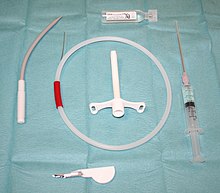
Several manufacturers market prepackaged cricothyrotomy kits, which enable one to use either a wire-guided percutaneous dilational (Seldinger) technique, or the classic surgical technique to insert a polyvinylchloride catheter through the cricothyroid membrane. The kits may be stocked in hospital emergency departments and operating suites, as well as ambulances and other selected pre-hospital settings.[60]
Tracheotomy
5 - Balloon cuff
Tracheotomy consists of making an incision on the front of the neck and opening a direct airway through an incision in the trachea. The resulting
Children
There are significant differences in airway anatomy and respiratory physiology between children and adults, and these are taken into careful consideration before performing tracheal intubation of any pediatric patient. The differences, which are quite significant in infants, gradually disappear as the human body approaches a mature age and body mass index.[64]
For infants and young children, orotracheal intubation is easier than the nasotracheal route. Nasotracheal intubation carries a risk of dislodgement of
Because the airway of a child is narrow, a small amount of glottic or tracheal swelling can produce critical obstruction. Inserting a tube that is too large relative to the diameter of the trachea can cause swelling. Conversely, inserting a tube that is too small can result in inability to achieve effective positive pressure ventilation due to retrograde escape of gas through the glottis and out the mouth and nose (often referred to as a "leak" around the tube). An excessive leak can usually be corrected by inserting a larger tube or a cuffed tube.[70]
The tip of a correctly positioned tracheal tube will be in the mid-trachea, between the
Newborn infants
Endotrachael suctioning is often used during intubation in newborn infants to reduce the risk of a blocked tube due to secretions, a collapsed lung, and to reduce pain.[7] Suctioning is sometimes used at specifically scheduled intervals, "as needed", and less frequently. Further research is necessary to determine the most effective suctioning schedule or frequency of suctioning in intubated infants.[7]
In newborns free flow oxygen used to be recommended during intubation however as there is no evidence of benefit the 2011 NRP guidelines no longer do.[71]
Predicting difficulty

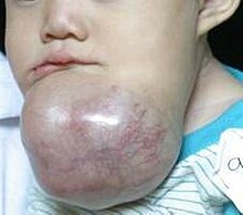
Tracheal intubation is not a simple procedure and the consequences of failure are grave. Therefore, the patient is carefully evaluated for potential difficulty or complications beforehand. This involves taking the medical history of the patient and performing a physical examination, the results of which can be scored against one of several classification systems. The proposed surgical procedure (e.g., surgery involving the head and neck, or bariatric surgery) may lead one to anticipate difficulties with intubation.[34] Many individuals have unusual airway anatomy, such as those who have limited movement of their neck or jaw, or those who have tumors, deep swelling due to injury or to allergy, developmental abnormalities of the jaw, or excess fatty tissue of the face and neck. Using conventional laryngoscopic techniques, intubation of the trachea can be difficult or even impossible in such patients. This is why all persons performing tracheal intubation must be familiar with alternative techniques of securing the airway. Use of the flexible fiberoptic bronchoscope and similar devices has become among the preferred techniques in the management of such cases. However, these devices require a different skill set than that employed for conventional laryngoscopy and are expensive to purchase, maintain and repair.[72]
When taking the patient's medical history, the subject is questioned about any significant
A detailed physical examination of the airway is important, particularly:[73]
- the range of motion of the cervical spine: the subject should be able to tilt the head back and then forward so that the chin touches the chest.
- the range of motion of the jaw (the temporomandibular joint): three of the subject's fingers should be able to fit between the upper and lower incisors.
- the size and shape of the lower jaw, looking especially for problems such as maxillary hypoplasia (an underdeveloped upper jaw), micrognathia (an abnormally small jaw), or retrognathia(misalignment of the upper and lower jaw).
- the thyromental distance: three of the subject's fingers should be able to fit between the Adam's apple and the chin.
- the size and shape of the tongue and palate relative to the size of the mouth.
- the teeth, especially noting the presence of prominent maxillary incisors, any loose or damaged teeth, or crowns.
Many classification systems have been developed in an effort to predict difficulty of tracheal intubation, including the
Complications
Tracheal intubation is generally considered the best method for airway management under a wide variety of circumstances, as it provides the most reliable means of oxygenation and ventilation and the greatest degree of protection against regurgitation and pulmonary aspiration.[2] However, tracheal intubation requires a great deal of clinical experience to master[83] and serious complications may result even when properly performed.[84]
Four anatomic features must be present for orotracheal intubation to be straightforward: adequate mouth opening (full range of motion of the temporomandibular joint), sufficient pharyngeal space (determined by examining the
Minor complications are common after laryngoscopy and insertion of an orotracheal tube. These are typically of short duration, such as sore throat, lacerations of the lips or
More serious complications include laryngospasm, perforation of the trachea or esophagus, pulmonary aspiration of gastric contents or other foreign bodies, fracture or dislocation of the cervical spine, temporomandibular joint or arytenoid cartilages, decreased oxygen content, elevated arterial carbon dioxide, and vocal cord weakness.[84] In addition to these complications, tracheal intubation via the nasal route carries a risk of dislodgement of adenoids and potentially severe nasal bleeding.[39][40] Newer technologies such as flexible fiberoptic laryngoscopy have fared better in reducing the incidence of some of these complications, though the most frequent cause of intubation trauma remains a lack of skill on the part of the laryngoscopist.[84]
Complications may also be severe and long-lasting or permanent, such as vocal cord damage, esophageal perforation and retropharyngeal abscess, bronchial intubation, or nerve injury. They may even be immediately life-threatening, such as laryngospasm and negative pressure pulmonary edema (fluid in the lungs), aspiration, unrecognized esophageal intubation, or accidental disconnection or dislodgement of the tracheal tube.[84] Potentially fatal complications more often associated with prolonged intubation or tracheotomy include abnormal communication between the trachea and nearby structures such as the innominate artery (tracheoinnominate fistula) or esophagus (tracheoesophageal fistula). Other significant complications include airway obstruction due to loss of tracheal rigidity, ventilator-associated pneumonia and narrowing of the glottis or trachea.[33] The cuff pressure is monitored carefully in order to avoid complications from over-inflation, many of which can be traced to excessive cuff pressure restricting the blood supply to the tracheal mucosa.[85][86] A 2000 Spanish study of bedside percutaneous tracheotomy reported overall complication rates of 10–15% and procedural mortality of 0%,[61] which is comparable to those of other series reported in the literature from the Netherlands[87] and the United States.[88]
Inability to secure the airway, with subsequent failure of oxygenation and ventilation is a life-threatening complication which if not immediately corrected leads to
One complication—unintentional and unrecognized intubation of the esophagus—is both common (as frequent as 25% in the hands of inexperienced personnel)[89] and likely to result in a deleterious or even fatal outcome. In such cases, oxygen is inadvertently administered to the stomach, from where it cannot be taken up by the circulatory system, instead of the lungs. If this situation is not immediately identified and corrected, death will ensue from cerebral and cardiac anoxia.
Of 4,460 claims in the
During the
Alternatives
Although it offers the greatest degree of protection against regurgitation and pulmonary aspiration, tracheal intubation is not the only means to maintain a patent airway. Alternative techniques for airway management and delivery of oxygen, volatile anesthetics or other breathing gases include the laryngeal mask airway, i-gel, cuffed oropharyngeal airway, continuous positive airway pressure (CPAP mask), nasal BiPAP mask, simple face mask, and nasal cannula.[98]
General anesthesia is often administered without tracheal intubation in selected cases where the procedure is brief in duration, or procedures where the depth of anesthesia is not sufficient to cause significant compromise in ventilatory function. Even for longer duration or more invasive procedures, a general anesthetic may be administered without intubating the trachea, provided that patients are carefully selected, and the
Airway management can be classified into closed or open techniques depending on the system of ventilation used. Tracheal intubation is a typical example of a closed technique as ventilation occurs using a closed circuit. Several open techniques exist, such as spontaneous ventilation, apnoeic ventilation or jet ventilation. Each has its own specific advantages and disadvantages which determine when it should be used.
Spontaneous ventilation has been traditionally performed with an inhalational agent (i.e. gas induction or inhalational induction using halothane or sevoflurane) however it can also be performed using intravenous anaesthesia (e.g. propofol, ketamine or dexmedetomidine). SponTaneous Respiration using IntraVEnous anaesthesia and High-flow nasal oxygen (STRIVE Hi) is an open airway technique that uses an upwards titration of propofol which maintains ventilation at deep levels of anaesthesia. It has been used in airway surgery as an alternative to tracheal intubation.[99]
History
- Tracheotomy
The earliest known depiction of a tracheotomy is found on two Egyptian tablets dating back to around 3600 BC.
The first detailed descriptions of tracheal intubation and subsequent
Despite the many recorded instances of its use since
In 1871, the German surgeon
- Laryngoscopy and non-surgical techniques
In 1854, a Spanish
In 1913, Chevalier Jackson was the first to report a high rate of success for the use of direct laryngoscopy as a means to intubate the trachea.
Between 1945 and 1952,
By the mid-1980s, the flexible fiberoptic bronchoscope had become an indispensable instrument within the pulmonology and anesthesia communities.
See also
Notes
- ^ a b c Benumof (2007), Ezri T and Warters RD, Chapter 15: Indications for tracheal intubation, pp. 371–8
- ^ PMID 16324990.
- ^ Advanced Trauma Life Support Program for Doctors (2004), Committee on Trauma, American College of Surgeons, Head Trauma, pp. 151–76
- ^ PMID 17460222.
- .
- ^ Doherty (2010), Holcroft JW, Anderson JT and Sena MJ, Shock & Acute Pulmonary Failure in Surgical Patients, pp. 151–75
- ^ PMID 26945780.
- S2CID 221389855.
- ^ Benumof (2007), Christodolou CC, Murphy MF and Hung OR, Chapter 17: Blind digital intubation, pp. 393–8
- ^ S2CID 6345531.
- ^ Benumof (2007), Berry JM, Chapter 16: Conventional (laryngoscopic) orotracheal and nasotracheal intubation (single lumen tube), pp. 379–92
- ISBN 978-1-4963-3700-9.
- ^ a b c Benumof (2007), Wheeler M and Ovassapian A, Chapter 18: Fiberoptic endoscopy-aided technique, p. 399-438
- PMID 35373840.
- S2CID 35038041.
- S2CID 21106547.
- ^ a b Benumof (2007), Hung OR and Stewart RD, Chapter 20: Intubating stylets, pp. 463–75
- PMID 12697606.
- PMID 15385401.
- S2CID 7977609.
- S2CID 13037753.
- ^ "Sheridan endotracheal tubes catalog" (PDF). Hudson RCI. 2002. Archived from the original (PDF) on 2011-04-09. Retrieved 2010-07-25.
- S2CID 18358131.
- S2CID 26644781.
- ^ a b US patent 5329940, Adair, Edwin L., "Endotracheal tube intubation assist device", published 1994-07-19, issued July 19, 1994
- ^ "Tracheostomy tube". Dictionary of Cancer Terms. National Cancer Institute. 2011-02-02.
- PMID 19247745.
- PMID 17375630. Archived from the original(PDF) on 2012-03-11. Retrieved 2010-08-30.
- S2CID 19119058.
- PMID 20749636.
- PMID 1168437.
- ^ a b Benumof (2007), Sheinbaum R, Hammer GB, Benumof JL, Chapter 24: Separation of the two lungs, pp. 576–93
- ^ a b c d Barash, Cullen and Stoelting (2009), Rosenblatt WH. and Sukhupragarn W, Management of the airway, pp. 751–92
- ^ a b c d e Miller (2000), Stone DJ and Gal TJ, Airway management, pp. 1414–51
- ^ Wolfe, T (1998). "The Esophageal Detector Device: Summary of the current articles in the literature". Salt Lake City, Utah: Wolfe Tory Medical. Archived from the original on 2006-11-14. Retrieved 2009-01-29.
- ^ Benumof (2007), Salem MR and Baraka A, Chapter 30: Confirmation of tracheal intubation, pp. 697–730
- PMID 8001220.
- .
- ^ PMID 3415064.
- ^ PMID 2321818.
- S2CID 39762822.
- ^ Benumof (2007), Hagberg CA and Benumof JL, Chapter 9: The American Society of Anesthesiologists' management of the difficult airway algorithm and explanation-analysis of the algorithm, pp. 236–54
- PMID 10941582.
- ^ Benumof (2007), Frass M, Urtubia RM and Hagberg CA, Chapter 25: The Combitube: esophageal-tracheal double-lumen airway, pp. 594–615
- ^ Benumof (2007), Suresh MS, Munnur U and Wali A, Chapter 32: The patient with a full stomach, pp. 752–82
- PMID 13749923.
- PMID 4593092.
- PMID 11983655.
- S2CID 18535821.
- S2CID 42387260.
- PMID 8467551.
- S2CID 16579238.
- PMID 16713784.
- S2CID 5459569.
- .
- ^ Benumof (2007), Melker RJ and Kost KM, Chapter 28: Percutaneous dilational cricothyrotomy and tracheostomy, pp. 640–77
- ^ Advanced Trauma Life Support Program for Doctors (2004), Committee on Trauma, American College of Surgeons, Airway and Ventilatory Management, pp. 41–68
- S2CID 33528267.
- ^ a b Benumof (2007), Gibbs MA and Walls RM, Chapter 29: Surgical airway, pp. 678–96
- S2CID 24568104.
- ^ PMID 11056749.
- PMID 2661159.
- S2CID 20209753.
- ^ Barash, Cullen and Stoelting (2009), Cravero JP and Cain ZN, Pediatric anesthesia, pp. 1206–20
- PMID 2297088.
- PMID 7856930.
- ]
- PMID 19690244.
- ^ Benumof (2007), Rabb MF and Szmuk P, Chapter 33: The difficult pediatric airway, pp. 783–833
- S2CID 24736705.
- PMID 21285656.
- S2CID 5827594.
- ^ Benumof (2007), Reed AP, Chapter 8: Evaluation and recognition of the difficult airway, pp. 221–35
- ^ Zadrobilek, E (2009). "The Cormack-Lehane classification: twenty-fifth anniversary of the first published description". Internet Journal of Airway Management. 5.
- S2CID 10561049.
- PMID 4027773.
- S2CID 12600824.
- S2CID 22147759.
- PMID 20554633.
- PMID 15459613.
- PMID 29761867.
- ^ Levitan (2004), Levitan RM, The limitations of difficult airway prediction in emergency airways, pp. 3–11
- PMID 17312190.
- ^ a b c d e f g Benumof (2007), Hagberg CA, Georgi R and Krier C, Chapter 48: Complications of managing the airway, pp. 1181–218
- PMID 15569386.
- ^ Benumof (2007), Pousman RM and Parmley CL, Chapter 44: Endotracheal tube and respiratory care, pp. 1057–78
- PMID 12740279.
- PMID 8760530.
- ^ PMID 11145768.
- PMID 15175215.
- PMID 9189188.
- PMID 9472186.
- S2CID 52931036.
- S2CID 6525904.
- PMID 32102726.
high-risk aerosol-producing procedures such as endotracheal intubation may put the anesthesiologists at high risk of nosocomial infections
- ^ "World Federation Of Societies of Anaesthesiologists - Coronavirus". www.wfsahq.org. 25 June 2020.
Anaesthesiologists and other perioperative care providers are particularly at risk when providing respiratory care and tracheal intubation of patients with COVID-19
- ^ "Clinical management of severe acute respiratory infections when novel coronavirus is suspected: What to do and what not to do" (PDF). World Health Organization. p. 4.
The most consistent association of in-creased risk of transmission to healthcare workers (based on studies done during the SARS outbreaks of 2002–2003) was found for tracheal intubation.
- ^ a b Benumof (2007), McGee JP, Vender JS, Chapter 14: Nonintubation management of the airway: mask ventilation, pp. 345–70
- PMID 28203745.
- S2CID 35712860.
- S2CID 34938843.
- PMID 3556136.
- ^ Bhishagratna (1907), Bhishagratna, Introduction, p. iv
- PMID 19567383.
- ^ Singer (1956), Galeni Pergameni C, De anatomicis administrationibus, pp. 195–207
- ^ PMID 4944603.
- ^ Longe (2005), Skinner P, Unani-tibbi
- ^ Shehata, M (2003). "The Ear, Nose and Throat in Islamic Medicine" (PDF). Journal of the International Society for the History of Islamic Medicine. 2 (3): 2–5.
- ^ Goodall, EW (1934). "The story of tracheostomy". British Journal of Children's Diseases. 31: 167–76, 253–72.
- ^ Habicot (1620), Habicot N, Question chirurgicale, p. 108
- PMID 14488739.
- ^ Trousseau, A (1852). "Nouvelles recherches sur la trachéotomie pratiquée dans la période extrême du croup". Annales de Médecine Belge et étrangère (in French). 1: 279–88.
- PMID 35376437.
- PMID 20319535.
- ^ Mackenzie (1888), Mackenzie M, The case of Emperor Frederick III, p. 276
- S2CID 221922284.
- ^ Radomski, T (2005). "Manuel García (1805–1906):A bicentenary reflection" (PDF). Australian Voice. 11: 25–41.
- PMID 18320839.
- PMID 20306068.
- S2CID 12259652.
- S2CID 72582327.
- ^ Jackson (1922), Jackson C, Instrumentarium, pp. 17–52
- S2CID 36279277.
- PMID 20775829.
- PMID 18956794.
- PMID 20770050.
- .
- .
- .
- .
- .
- ^ "History of endoscopes. Volume 2: Birth of gastrocameras". Olympus Corporation. 2010.
- ^ "History of endoscopes. Volume 3: Birth of fiberscopes". Olympus Corporation. 2010.
- S2CID 33586314.
- PMID 638831.
References
- Barash, PG; Cullen, BF; Stoelting, RK, eds. (2009). Clinical Anesthesia (6th ed.). Philadelphia: Lippincott Williams & Wilkins. ISBN 978-0-7817-8763-5.
- Benumof, JL, ed. (2007). Benumof's Airway Management: Principles and Practice (2nd ed.). Philadelphia: Mosby-Elsevier. ISBN 978-0-323-02233-0.
- Bhishagratna, KL, ed. (1907). Sushruta Samhita, Volume1: Sutrasthanam. Calcutta: Kaviraj Kunja Lal Bhishagratna.
- ISBN 978-1-880696-31-6.
- Doherty, GM, ed. (2010). Current Diagnosis & Treatment: Surgery (13th ed.). New York: McGraw-Hill Medical. ISBN 978-0-07-163515-8.
- Habicot, N (1620). Question chirurgicale par laquelle il est démonstré que le chirurgien doit assurément practiquer l'operation de la bronchotomie, vulgairement dicte laryngotomie, ou perforation de la fluste ou du polmon (in French). Paris: Corrozet.
- ISBN 978-1-4326-6305-6. Archived from the original on 2010-02-19. Retrieved 2010-08-03.
- Levitan, RM (2004). The Airway Cam Guide to Intubation and Practical Emergency Airway Management (1st ed.). Wayne, Pennsylvania: Airway Cam Technologies. ISBN 978-1-929018-12-3.
- Longe, JL, ed. (2005). "The Gale Encyclopedia of Alternative Medicine: L-R". The Gale Encyclopedia of Alternative Medicine, Volume 4: S–Z (2nd ed.). Farmington Hills, Michigan: Gale Cengage. ISBN 978-0-7876-7424-3.
- Mackenzie, M (1888). The case of Emperor Frederick III.: full official reports by the German physicians and by Sir Morell Mackenzie. New York: Edgar S. Werner.
- Miller, RD, ed. (2000). Anesthesia, Volume 1 (5th ed.). Philadelphia: Churchill Livingstone. ISBN 978-0-443-07995-5.
- ISBN 978-0-19-924016-6.
External links
- Video of endotracheal intubation using C-MAC D-blade and bougie used as introducer.
- Videos of direct laryngoscopy recorded with the Airway Cam (TM) imaging system
- Examples of some devices for facilitation of tracheal intubation
- Free image rich resource explaining various types of endotracheal tubes
- Tracheal intubation live case 2022
