
Brenner tumour
| Brenner tumor | |
|---|---|
gross image). | |
| Specialty | Gynaecology, oncology |
Brenner tumours are an uncommon subtype of the
They are most frequently found incidentally on
Presentation
On gross pathological examination, they are solid, sharply circumscribed and pale yellow-tan in colour. 90% are unilateral (arising in one ovary, the other is unaffected). The tumours can vary in size from less than 1 centimetre (0.39 in) to 30 centimetres (12 in). Borderline and malignant Brenner tumours are possible but each are rare.
Diagnosis


The coffee bean nuclei are the nuclear grooves exceptionally pathognomonic to the sex cord stromal tumour, the ovarian granulosa cell tumour, with the fluid-filled spaces Call–Exner bodies between the granulosa cells.[4][5]
Similar conditions
Transitional cell carcinoma is an even rarer entity, in which neoplastic transitional epithelial cells similar to transitional cell carcinoma of the bladder are seen in the ovary, without the characteristic stromal/epithelial pattern of a Brenner tumour.
Histologically, Leydig cell tumours of the testes and ovarian stromal Leydig cell tumours (ovarian hyperandrogenism and virilization) both have characteristic Reinke crystals. The same crystals were also noted under high-power view in Brenner tumours.[6]
Eponym
It is named for
Additional images
-
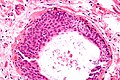 Walthard cell nest, the entity Brenner tumours are thought to arise from. H&E stain.

References
- PMID 16758686.
- PMID 16998096.
- PMID 2021324.
- ^ "Pathology Thread". University of Virginia Medical School. Archived from the original on 4 February 2006.
- PMID 9990262.
- PMID 30377407.
- PMID 903146.
- ISBN 978-1-85070-040-1.
External links
- "Brenner tumour". Medcyclopaedia. GE. Archived from the original on 2012-02-05.
- Histology at University of Utah