
Liposarcoma
| Liposarcoma | |
|---|---|
 | |
| Histopathology of liposarcoma, H&E stain:[1] - | |
| Specialty | Dermatology, general surgery oncology |
| Symptoms | Lump under skin, pain, swelling, organ dysfunction |
Liposarcomas are the most common subtype of soft tissue
All liposarcomas consist of at least some cells that bear a resemblance to fat cells when examined for their
While liposarcoma forms are classified as being aggressive and
Etymology
"fatty tumor" (plural lipomata), 1830, medical Latin, from Greek lipos "fat" (n.), from PIE root *leip- "to stick, adhere", also used to form words for "fat", + -oma.
1650s, "fleshy excrescence", (plural liposarcomata), Medical Latin, from Latinized form of Greek sarkoma "fleshy substance" (Galen), from sarkoun "to produce flesh, grow fleshy", from sarx (genitive sarkos) "flesh", + -oma.
Forms of liposarcomas

Liposarcomas are generally large tumors (>10 cm) but can be of almost any size. They occur mainly in adults with only 0.7% of cases occurring in those <16 years old.[5] In adults, liposarcomas occur predominantly in and after middle-age.[8] The very rare cases occurring in children and adolescents are diagnosed predominantly as being the myxoid liposarcoma form.[5]
The five liposarcoma forms must be distinguished not only from each other but also from certain other soft tissue tumors. These other tumors along with some of their distinguishing histopathologic features are: 1) dysplastic lipomas (i.e. benign humors that have sites of tissue
Atypical lipomatous tumor/well-differentiated liposarcoma
Together, atypical lipomatous tumors (ALTs) and well-differentiated liposarcomas (WDLs) account for 40–45% of all liposarcomas.
Presentation

ALTs and WDLs are considered virtually identical tumors except that by definition ALTs designate tumors that develop in the arms or legs while WDLs designate tumors that develop in less surgically accessible sites such as the deep, centrally-located soft tissues of the
Pathology
Histopathologically, ALT/WDL tumors are divided into adipocytic/lipoma-like, sclerosing, and inflammatory variants with adipocyte/lipoma-like being the most common. Adipocytic/lipoma-like ALT/WDL tumors consist of lobules of mature fat cells variably intersected with irregular fibrous
Genetics
The neoplastic cells in ALT/WDL tumors contain one or more extra ring-shaped
Diagnosis
The diagnosis of ALT/WDL tumors is made based on the features of their clinical presentations, histopathology, and genetic findings. In particular, detection in the ALT/WDL tumor cells of an overexpressed MDM2 or CDK4 gene or the presence of either the specific ALT/WDL-associated sSMC or giant marker chromosome (as defined by next generation DNA sequencing, comparative genomic hybridization,[18] and/or highly specialized cytogenetic G banding analyses[19]) strongly supports the diagnosis of ALT/WDL or dedifferentiated liposarcoma. The clinical presentation and histopathology differences between the latter two liposarcoma forms usually help distinguish between them.[8]
Treatment and prognosis
ALT/WDL tumors are treated by radical surgical resection to remove all tumor neoplastic tissues. However, these tumors recur locally in 30–50% of cases. Recurrences occur most often in tumors located in less accessible sites such those in the retroperitoneum, mediastinum, and spermatic cord. These less surgically assessible tumors tend to recur repeatedly and ultimately may cause death due to their injurious effects on vital organs. While ALT/WDL tumors have very little potential to
Novel therapies
The novel therapies of ALT/WDL are the same as those listed in the Novel therapies section of Dedifferentiated liposarcoma.[citation needed]
Dedifferentiated liposarcoma
Dedifferentiated liposarcomas are malignant tumors which in ~10% of cases develop in an existing atypical lipomatous tumor/well-differentiated liposarcoma (ALT/WDL) tumor or at the site were an ALT/WPL tumor was surgically removed. Individuals with a de novo diagnosis of this tumor may have had an ALT/WDL that progressed to a dedifferentiated liposarcoma but went undetected because it developed asymptomatically in a highly sequestered site such as the retroperitoneum or abdominal cavity. Many of the dedifferentiated liposarcoma tumors' clinical and genetic features are similar to those found in ALT/WDL tumors.[8]
Presentation
Dedifferentiated lipoosarcomas (DDL) occur most frequently in middle-aged and older adults with a peak incidence in their sixth to eighth decades.
Pathology
The
Genetics
The neoplastic cells in both DDL and ALT/WDL carry similar
Diagnosis
The histopathological of DDL is often insufficiently clear to make a firm diagnosis. However, the diagnosis of DDL is supported in individuals: whose tumors contain ALT/WDL admixed with DDL histological components; with histories of having a prior ALT/WDL;[8] or who present with a retroperitoneal liposarcoma (DDL constitutes ~57% of all retroperitoneal liposarcomas). DDL tumors only rarely (<1% of cases) present as superficial skin tumors;[8] are almost 5 times less likely than ALT/WDL to occur in the eye socket;[14][21] and are extremely rare in children.[5] Detection of tumor cell MDM2 amplification is the diagnostic gold standard in distinguishing WDL from lipomas, dysplastic lipomas, atypical spindle cell sarcomas, pleomorphic lipomas, and solitary fibrous tumors.[8] Alternately, detection in the tumor cells of an overexpressed CDK4 gene or the presence of either the specific ALT/WDL-associated sSMCs or giant marker chromosome strongly support the diagnosis of DDL or ALT/WDL.[18][19] The clinical presentation, histopathology, and gene differences (e.g. tumor cell overexpression of the cJUN gene strongly favors the diagnosis of DDL over ATL/WDL) between the latter two liposarcoma forms usually help distinguish between them.[8]
Treatment and Prognosis
Complete surgical resection is usually the recommended first-line treatment for localized DDL tumors.
Retroperitoneal DDL is the most common, surgically unaccessible and serious form of DDL: it has a recurrence rate of 66% and a five-year overall survival rate of 54%.[31] The primary treatment option for retroperitoneal DDL is surgical resection. A phase III clinical trial found little difference in the results of radiation therapy followed by surgical resection compared to surgical resection alone in the treatment of retroperitoneal DDL.[6] In other phase III clinical trials, DDL patients with inaccessible retroperitoneal and/or metastatic tumors were treated with front-line chemotherapy comparing doxorubicin to doxorubicin plus ifosfamide or doxorubicin to gemcitabine plus docetaxel. Other studies have likewise examined the value of various chemotherapy regimens. These studies often found little difference in the overall survival times in their comparisons but did show some improvements in progression-free survival and other clinical parameters. Based on these studies, a recommended first-line therapy for retroperitoneal and other surgically inassessible or metastatic DDL tumors is treatment with an anthracycline-based chemotherapy regimen or, in tumor-resistant or relapsed cases, eribulin chemotherapy. A review conducted in 2020 reported median survival times for low histopathological grade and high histopathological grade DDL to be 113 months and 48 months, respectively.[32] Further studies are needed to provide evidence on the efficacies of radiotherapy, chemotherapy, and novel therapies in all the varieties of DDL.[33]
Novel therapies
Several novel therapy regimens for DDL and the more aggressive or otherwise problematic cases of ALT/WDL are currently undergoing
Myxoid liposarcoma
Presentation
Myxoid liposarcoma (MLS), which includes a type of liposarcoma termed round cell liposarcoma,[39] represents ~30% of all liposarcomas. It has a peak incidence in individuals' fourth and fifth decades with a male predominance in most studies. While uncommon in children and adolescents, MLS is the most common liposarcoma form diagnosed in these age groups. MLS typically presents as a large (1 to 39 cm; average 12 cm), mobile, well-circumscribed, painless mass that developed from 1 week to 15 years prior to diagnosis. MLS tumors are located in deep-seated soft tissues of the thighs (65–80% of cases), lower legs (10–15% of cases), retroperitoneum (8% of cases), and arms (5% of cases). In about one-third of cases, these tumors metastasize to other soft tissue sites (e.g. retroperitoneum, thorax, or other extremity), skeletal bone, and/or lung. Individuals may present with these metastasis, particularly those in bone; it has been recommended that patients should be tested at presentation for bone metastasis by medical imaging, including X-rays, CT scans, and/or magnetic resonance imaging.[40]
Pathology
Histopathologic analyses of MLS (see Figs. 3 and 4 in the below Histopathology of liposarcomas section) reveals cells scattered throughout a myxoid matrix (i.e. a connective tissue background that appears more blue or purple than the red color of normal connective tissue when these tissues are properly prepared, H&E stained, and viewed microscopically). These cells are lipoblasts, some of which are signet ring-shaped (a shape suggesting that the cell may be neoplastic), oval-shaped, or round-shaped.[40] MLS tumors may be hypercellular and contain solid sheets of round cells that comprise at least 5% of all cells or low cellularity populated with cells that have bland nuclei and <5% round cells in a background of curving capillaries resembling a chicken-wire pattern. Tumors that contain at least 5% round cells are classified as high-grade while those with <5% round cells are classified as low-grade.[39] High-grade MLS tumors typically take a more aggressive clinical course than low-grade MLS tumors.[40]
Genetics
MLS tumor cells are virtually defined by their expression of a FUS-DDIT3
Diagnosis
Low-grade and intermediate-grade MLS tumors can be identified histologically by their classic morphology of distinctive chicken-wire vasculature scattered throughout a myxoid stroma. However, high-grade MLS tumors can be difficult to distinguish from other round cell neoplasms, particularly high grade MLS tumors that consist of diffuse cell and/or pure round cell morphology to such an extent as to obscure this classic vascular-myxoid pattern. Detection of a DDIT3 gene rearrangements with the FUS or EWSR1 gene by in situ hybridization or immunohistochemistry or the RNA fusion transcripts of these genes by real-time polymerase chain reactions confirms the diagnosis of high-grade as well as ambiguous cases of low-grade or intermediate-grade MLS tumors.[44]
Treatment and prognosis
MLS has typically been treated by surgical resection but may require more radical interventions, e.g. limb amputation may be needed when a limb's neurovascular bundle is compromised. The post-surgical risk of recurrence within 3 years after surgery has been reported to be ~15% when not all tumor is removed and ~10% when tumor removal is complete.[40] The addition of radiotherapy to surgical resection has improved the local control of MLS tumors and has been recommended to treat unresectable and recurrent MLS.[45] However, further studies are needed to determine the value of radiotherapy in treating the various varieties of MLS.[40] Chemotherapy regimens using ifosfamide, an anthracycline such as daunorubicin, dacarbazine, and/or trabectedin have been found useful: a phase III clinical trial showed progression-free survival times in MLS patients treated with trabectedin or dacarbazine to be 5.6 and 1.5 months, respectively. In 2015 the Food and Drug Administration approved trabectedin for use in unresectable and metastatic liposarcomas.[citation needed]
Overall, the 10-year survival rate of MLS individuals has been 77%, a survival rate appreciably longer than other liposarcoma forms. Compared to low-risk MLS, high-risk MLS (risk defined by tumor round cell content and/or other unfavorable prognostic indicators) is associated with increased rates of metastasis and therefore a shorter survival time. Increased tumor size (≥ 10 cm) is strongly associated with a higher grade MLS and therefore a shorter survival time. Other factors that have been associated with unfavorable outcomes in MLS include presence of tumor necrosis, age >45 years, P53 gene overexpression,[40] and male gender.[46] The round cell form of myxoid liposarcomas also appears to have a relatively poor prognosis: in various retrospective reviews, myxoid liposarcoma was usually found to be low-grade and therefore relatively responsive to chemotherapy whereas high grade (i.e. round cell) myxoid lipsarcoma had higher rates of metastasis, behaved more aggressively, and did not respond well to chemotherapy.[40] It is important to note, however, that almost all cases of myxoid liposarcomas in pediatric patients have had excellent prognoses.[45]
Novel therapies
A
Pleomorphic liposarcoma
Presentation
Pleomorphic liposarcomas (PLS), which account for 5% to 10% of all liposarcoma cases,
Pathology
The histopathology of PLS tumors often consists of areas resembling myxoid liposarcoma
Genetics
PLS neoplastic cells contain various gene and chromosome abnormalities: the
Diagnosis
The diagnosis of PLS depends on its presentation, histopathology, and genetics. The histopathology of PLS often closely resembles that of myxofibrosarcoma but is distinguished from that tumor by its content of pleomorphic lipoblasts.[58]
Treatment and prognosis
Radical surgical resection is the main treatment for PLS; it is also an important palliative intervention to relieve symptoms due to the compression of organs and tissues. Surgery may require removal of an entire compressed organ such as the kidney or colon. Regardless of this surgery, however, local recurrence rates are very high. The uses of chemotherapy and/or radiotherapy in conjunction with radical surgery have not been shown to prolong survival and are regarded as controversial interventions.[55] The National Comprehensive Cancer Network recommends treatment for individuals with high-risk localized PLS by complete surgical resection, when feasible, combined with radiation therapy. Individuals with metastatic disease have been treated with chemotherapy (e.g. doxorubicin plus ifosfamide or eribulin) similar to the regimens used for dedifferentiated liposarcoma (see above section on the treatment of this liposarcoma type)[6] About 20% of PLS tumors metastasize to distant sites, the most common of which are lung (82% of metastases), liver (18% of metastases), and bone or pancreas (18% of metastases). PLS survival rates at 1, 3, and 5 years are reported to be 93%, 75%, and 29%, respectively. Tumors located in the center position of the trunk, larger than 10 cm in size, deeply seated, or containing areas of necrosis have worse prognoses.[55]
Myxoid pleomorphic liposarcoma
Myxoid pleomorphic liposarcoma (originally termed pleomorphic myxoid liposarcoma[62]) was first described in a large 2009 study of the liposarcomas.[63] While initially regarded as a possible variant of the myxoid liposarcomas with pleomorphic features, the World Health Organization (2020) classified it as a new and distinct form of the liposarcomas. This classification was based on findings that the myxoid pleomorphic liposarcomas, while having histopathological features that were similar to myxoid liposarcomas, had clinical and, most importantly, critical genetic and molecular features that differed from the myxoid as well as the other three liposarcoma forms.[5]
Presentation
Myxoid pleomorphic liposarcoma (MPL) is an exceptionally rare and highly aggressive form of the liposarcomas that develops in children, adolescents,[5] young adults,[6] and, in a more recent study, individuals >50 years old.[62] MPL tumors present as deep soft-tissue masses that are often located in the mediastinum[44] and, less often, the extremities, head and neck, abdominal cavity, or trunk.[6] At least two case of MPL have presented in individuals with the Li–Fraumeni syndrome, an inherited genetic disorder that predisposes individuals to develop various cancers.[58][64][65]
Pathology
On histopathologic analyses, MPL tumors consist of areas resembling conventional myxoid liposarcoma; these areas, which represent 30–50% of the total tumor areas, have an abundant myxoid matrix, a well-developed capillary vasculature, bland cells that are round and/or slightly spindle-shaped, vacuolated lipoblasts, and multinucleated cells shaped like small flowers. However, these areas also contain a scattering of highly pleomorphic cells that show greater degrees of nuclear enlargement and irregularity than the cells seen myxoid liposarcoma tumors. Other areas of MPL tumors are more cellular and consist of rapidly growing and highly pleomorphic lipoblasts.[62]
Genetics
The neoplastic cells in MPL do not express the FUS-DDIT3 or EWSR1-DDIT3 fusion genes that are expressed by the neoplastic cells in >95% or <5%, respectively, of myxoid fibrosarcoma cases.
Diagnosis
The diagnosis of MPL depends on its tumors clinical presentation, histopathological resemblance to myxoid liposarcoma, and, most critically, absence of the FUS-DDIT3 sn EWSR1-DDIT3 fusion genes in its neoplastic cells.[62][6]
Treatment and prognosis
While individuals with MPL have been treated with surgical resection to remove their tumors,[64][65][6][66] a 2021 review found that there were no consensus recommendations for the standard of care for MPL with respect to radiation and chemotherapy regimens (when used either alone or combined with surgery) for treating these tumors.[6]
Histopathology of liposarcomas
-
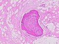 Fig. 1 Micrograph of bone formation in a liposarcoma tumor
 -
 Fig. 2 Micrograph of a dedifferentiated liposarcoma tumor
 -
 Fig. 3 Lower-power micrograph of myxoid liposarcoma tumor
 -
 Fig. 4 Higher-power micrograph of myxoid liposarcoma tumor


.jpg)
.JPG)
Medical imaging
In myxoid liposarcoma, it shows low signal intensity mass with high signal intensity foci on T1-weighted MRI images. The mass shows high signal intensity on T2-weighted images. This is because it contains predominantly mucoid substance (accounts for low signal intensity on T1) and small amount of mature fat (accounts for high signal intensity on T1).[68] The mass is well-defined, lobulated, multiloculated, or oval in shape without any infiltration into surrounding structures.[68]
-
![Fig. 5 Ultrasonography of a liposarcoma with high-echo areas reflected from its lipomatous matrix and low-echo areas reflected from its non-lipomatous areas.[69]](//upload.wikimedia.org/wikipedia/commons/thumb/a/a9/Scrotal_ultrasonography_of_liposarcoma.jpg/120px-Scrotal_ultrasonography_of_liposarcoma.jpg) Fig. 5Ultrasonography of a liposarcoma with high-echo areas reflected from its lipomatous matrix and low-echo areas reflected from its non-lipomatous areas.[69]
Fig. 5Ultrasonography of a liposarcoma with high-echo areas reflected from its lipomatous matrix and low-echo areas reflected from its non-lipomatous areas.[69] -
![Fig. 6 Ultrasonography of a liposarcoma mimicking a lipoma. This homogeneous high-echoic mass has the same appearance as a lipoma.[69]](//upload.wikimedia.org/wikipedia/commons/thumb/3/34/Scrotal_ultrasonography_of_liposarcoma_mimicking_a_lipoma.jpg/120px-Scrotal_ultrasonography_of_liposarcoma_mimicking_a_lipoma.jpg) Fig. 6Ultrasonography of a liposarcoma mimicking a lipoma. This homogeneous high-echoic mass has the same appearance as a lipoma.[69]
Fig. 6Ultrasonography of a liposarcoma mimicking a lipoma. This homogeneous high-echoic mass has the same appearance as a lipoma.[69] -
 Fig. 7MRI of myxoid liposarcoma of high grade, in the left axillary regionof 40-year-old man, highlighted by its white color, in this horizontal section of the tumor.
![Fig. 5 Ultrasonography of a liposarcoma with high-echo areas reflected from its lipomatous matrix and low-echo areas reflected from its non-lipomatous areas.[69]](/File:Scrotal_ultrasonography_of_liposarcoma.jpg)
![Fig. 6 Ultrasonography of a liposarcoma mimicking a lipoma. This homogeneous high-echoic mass has the same appearance as a lipoma.[69]](/File:Scrotal_ultrasonography_of_liposarcoma_mimicking_a_lipoma.jpg)

Society and culture
Notable cases
- Chad Brown (1961–2014), a poker player, died from liposarcoma
- Richard Feynman (1918–1988), a theoretical physicist, died following surgery to address the disease.
- Rob Ford (1969–2016), former Toronto mayor and Toronto city councillor, died of pleomorphic liposarcoma.
- Hokie Gajan (1959–2016), former running back for the New Orleans Saints and radio color commentator for the team, died from liposarcoma.
- Charlie Davies (born 1986), former soccer player for the Philadelphia Union of Major League Soccer, diagnosed with liposarcoma in 2016.
- Mark Strand (1934–2014), former US Poet Laureate and Pulitzer Prize-winner, died from liposarcoma.
See also
- Lipoma
- The Wendy Walk, not-for-profit organization whose mission is to raise funds and awareness for sarcomas, including liposarcoma
References
- ^ Susan Potterveld; Michael R. Clay. "Liposarcoma". PathologyOutlines. Topic Completed: November 2017. Minor changes: May 2023
- S2CID 229688954.
- PMID 10982304.
- ^ Bell, Teresa (October 2012). "What is Liposarcoma?". The Liddy Shriver Sarcoma Initiative. Retrieved 2015-04-22.
- ^ S2CID 235678096.
- ^ PMID 33659920.
- PMID 21274402.
- ^ S2CID 73725589.
- S2CID 51616357.
- PMID 34200924.
- ^ S2CID 27183708.
- ^ S2CID 232299293.
- PMID 30397612.
- ^ S2CID 222143763.
- ^ "MDM2 MDM2 proto-oncogene [Homo sapiens (Human)] - Gene - NCBI".
- ^ "CDK4 cyclin dependent kinase 4 [Homo sapiens (Human)] - Gene - NCBI".
- ^ PMID 27073568.
- ^ PMID 32399795.
- ^ PMID 32536972.
- ^ Gebhardt, M; Buecker, PJ (2004). "Liposarcoma". ESUN.
- ^ S2CID 216594976.
- S2CID 3829557.
- PMID 32190631.
- S2CID 207094296.
- S2CID 218680437.
- ^ "MDM2 MDM2 proto-oncogene [Homo sapiens (Human)] – Gene – NCBI".
- ^ "CDK4 cyclin dependent kinase 4 [Homo sapiens (Human)] – Gene – NCBI".
- PMID 29279323.
- ^ "YEATS4 YEATS domain containing 4 [Homo sapiens (Human)] – Gene – NCBI".
- S2CID 59341549.
- PMID 34150840.
- PMID 32774015.
- S2CID 235701424.
- ^ "Sarcoma Alliance for Research through Collaboration".
- ^ a b "SARC041: Phase 3 Randomized Double-Blind Study of Abemaciclib Versus Placebo in Patients with Advanced Dedifferentiated Liposarcoma". 23 July 2021.
- ^ "A Randomized Multicenter Phase 3 Study of Milademetan Versus Trabectedin in Patients with Dedifferentiated Liposarcoma". 16 July 2021.
- ^ "Milademetan".
- ^ "A Randomized Multicenter Phase 3 Study of Milademetan Versus Trabectedin in Patients with Dedifferentiated Liposarcoma". 16 July 2021.
- ^ PMID 30855853.
- ^ S2CID 208954696.
- ^ "DDIT3 DNA damage inducible transcript 3 [Homo sapiens (Human)] – Gene – NCBI".
- ^ "FUS FUS RNA binding protein [Homo sapiens (Human)] – Gene – NCBI".
- ^ "EWSR1 EWS RNA binding protein 1 [Homo sapiens (Human)] – Gene – NCBI".
- ^ S2CID 232247392.
- ^ S2CID 73513220.
- S2CID 216560584.
- ^ "Efatutazone dihydrochloride".
- ^ "A Phase II Study of the Peroxisome Proliferator-Activated Receptor Gamma Agonist, Efatutazone in Patients with Previously Treated, Unresectable Myxoid Liposarcoma". 16 August 2021.
- ^ "A Phase II Study on Trabectedin in Combination with PPARg Agonist Pioglitazone in Patients with Round Cell Myxoid Liposarcomas or Dedifferentiated G1 and G2 Liposarcomas with Stable Disease After a Monotherapy with Trabectedin. (TRABEPIO)". 10 March 2021.
- ^ "A Phase 2, Single Arm, Multi Center Trial Evaluating the Efficacy of the COmbination of Sirolimus and cYclophosphamide in Metastatic or Unresectable Myxoid Liposarcoma and chOndrosarcoma". 4 June 2021.
- ^ Luo, Zhiguo (13 July 2020). "A Single Arm, Multi Centers, Phase II Study of Sintilimab, Doxorubicin and Ifosfamide at First-line Treatment of Soft Tissue Sarcoma Including Undifferentiated Pleomorphic Sarcoma, Synovial Sarcoma, Myxoid Liposarcoma and De-differentiated Liposarcoma".
- PMID 32002290.
- PMID 32540953.
- ^ "A Phase 2 Single Arm Open-Label Clinical Trial of ADP-A2M4 SPEAR™ T Cells in Subjects with Advanced Synovial Sarcoma or Myxoid/Round Cell Liposarcoma". 18 June 2021.
- ^ PMID 29465602.
- ^ PMID 33802620.
- PMID 28528729.
- ^ S2CID 75135591.
- ^ "Pleomorphic liposarcoma".
- PMID 30018380.
- PMID 33363928.
- ^ S2CID 235614580.
- S2CID 21863759.
- ^ S2CID 203568504.
- ^ S2CID 29704574.
- S2CID 232273351.
- ^ Rohit Sharma; Frank Gaillard; et al. "Lipoma". Radiopaedia. Retrieved 2018-09-27.
- ^ PMID 10903690.
- ^ ISBN 978-953-307-947-9. under the CC-BY-3.0 license.