Cystadenocarcinoma
Appearance
| Ovarian Cystadenocarcinoma | |
|---|---|
| Other names | cystadenoma carcinoma |
 | |
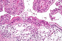
| Intermediate magnification micrograph of a low malignant potential (LMP) mucinous ovarian tumour. H&E stain.
The micrograph shows: Simple mucinous epithelium (right) and mucinous epithelium that pseudo-stratifies (left - diagnostic of a LMP tumour). Epithelium in a frond-like architecture is seen at the top of image. | |
Liposomal doxorubicin, etoposide, topotecan, gemcitabine, docetaxel, vinorelbine, ifosfamide, fluorouracil, melphalan, altretamine, bevacizumab, olaparib, rucaparib, niraparib, mesna . | |
Cystadenocarcinoma is a malignant form of a
ovaries,[1] where pseudomucinous and serous types are recognized. Similar tumor histology has also been reported in the pancreas, although it is a considerably rarer entity representing 1–1.5% of all Pancreatic cancer.[2][3]
A cystadenocarcinoma contains complex multi-loculated cyst but with exuberant solid areas in places. It usually presents with omental metastases which cause fluid accumulation in the peritoneal cavity (ascites). Cystadenocarcinomas can be classified into serous cystadenocarcinomas and mucinous cystadenocarcinomas.[citation needed]
Serous cystadenocarcinomas
Among the ovarian tumours, serous tumours are most common, having a variegated appearance. Bilateral presentation is common with serous cystadenocarcinoma.[4]
See also
References
- ^ "Ovarian papillary cystadenocarcinoma". Female Genital Pathology, WebPath, The Internet Pathology Laboratory for Medical Education. Eccles Health Sciences Library, The University of Utah. Retrieved 2009-03-23.
- ISBN 978-0-470-03055-4.
- PMID 19459016.
- ISBN 978-93-5025-369-4.[page needed]
