
Renal oncocytoma
| Renal oncocytoma | |
|---|---|
 | |
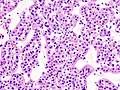
| Micrograph of a renal oncocytoma. |
A renal
Signs and symptoms

Renal oncocytomas are often asymptomatic and are frequently discovered by chance on a
Pathophysiology
Renal oncocytoma is thought to arise from the
Histologic appearance
This section needs expansion. You can help by adding to it. (January 2011) |
An oncocytoma is an
Diagnosis
In
The main differential diagnosis of renal oncocytoma is chromophobe renal cell carcinoma oncocytic variant, which like the renal oncocytoma has eosinophilic cytoplasm, but has perinuclear clearing and, typically, some degree of nuclear atypia.
Immunohistochemical profile
- Cytokeratin AE1/AE3 +
- Vimentin -
- EMA+
- CD10+/-
- CK7+/- parcellaire
- CK20-
- CK19-
- Racemase) -
- S100 +
- c-kit (CD117) +
- Cadherin-E +
- TFE3 -
- CAM5.2+
Treatment
Renal oncocytoma is considered benign, cured by nephrectomy. There are some familial cases in which these tumors are multicentric rather than solitary.[4] However, they may be resected to exclude a malignant tumor, e.g. renal cell carcinoma.
Prognosis

The overall five-year survival rate has been estimated to be 63%, with 100% disease-specific survival.[5]
Additional images
-
 Micrograph of a renal oncocytoma. H&E stain.
 -
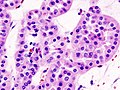
 Micrograph of a renal oncocytoma. H&E stain.
 -
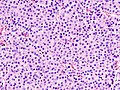
 Micrograph of a renal oncocytoma. H&E stain.
 -
 Micrograph of a renal oncocytoma. H&E stain.
 -
 Micrograph of a renal oncocytoma. H&E stain.
 -
 Micrograph of chromophobe RCC oncocytic variant, the main differential diagnosis of renal oncocytoma.


.jpg)
.jpg)
.jpg)

See also
References
- S2CID 19955625.
- ^ "Atlas of Genetics and Cytogenetics in Oncology and Haematology - Thyroid:oncocytic tumors". Retrieved 2009-02-01.
- ^ [Renal Neoplasms: An Update on Immunohistochemical and Histochemical Features http://www.dako.com/08066_12may10_webchapter26.pdf]
- ^ Robbins pathology, page 1015[full citation needed]
- S2CID 19390518.