
Hand
| Hand | |
|---|---|
 Back of a human's left hand | |
 Front of a human's left hand | |
| Details | |
| Vein | Dorsal venous network of hand |
| Nerve | Ulnar, median, radial nerves |
| Identifiers | |
| Latin | manus |
| MeSH | D006225 |
| TA98 | A01.1.00.025 |
| TA2 | 148 |
| FMA | 9712 |
| Anatomical terminology | |
A hand is a
Some evolutionary
The human hand usually has five digits:
Fingers contain some of the densest areas of nerve endings in the body, and are the richest source of
Among humans, the hands play an important function in
Structure
Many
The hand is located at the distal end of each arm. Apes and monkeys are sometimes described as having four hands, because the toes are long and the hallux is opposable and looks more like a thumb, thus enabling the feet to be used as hands.
The word "hand" is sometimes used by evolutionary anatomists to refer to the appendage of digits on the forelimb such as when researching the homology between the three digits of the bird hand and the dinosaur hand.[2]
An adult human male's hand weighs about a pound.[9]
Areas

Areas of the human hand include:
- The palm (volar), which is the central region of the anterior part of the hand, located superficially to the dermal papillae to increase friction, such as are also present on the fingers and used for fingerprints.
- The opisthenar area (dorsal) is the corresponding area on the posterior part of the hand.
- The heel of the hand is the area anteriorly to the bases of the metacarpal bones, located in the proximal part of the palm. It is the area that sustains most pressure when using the palm of the hand for support, such as in handstand.
There are five
can be folded over the palm which allows the grasping of objects. Each finger, starting with the one closest to the thumb, has a colloquial name to distinguish it from the others:- index finger, pointer finger, forefinger, or 2nd digit
- middle finger or long finger or 3rd digit
- ring finger or 4th digit
- little finger, pinky finger, small finger, baby finger, or 5th digit
The thumb (connected to the first metacarpal bone and trapezium) is located on one of the sides, parallel to the arm. A reliable way of identifying human hands is from the presence of opposable thumbs. Opposable thumbs are identified by the ability to be brought opposite to the fingers, a muscle action known as opposition.
Bones


The
Because
There are numerous
The articulations are:
- interphalangeal articulations of hand (the hinge jointsbetween the bones of the digits)
- metacarpophalangeal joints (where the digits meet the palm)
- intercarpal articulations(where the palm meets the wrist)
- wrist (may also be viewed as belonging to the forearm).
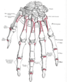
Arches

Red: one of the oblique arches
Brown: one of the longitudinal arches of the digits
Dark green: transverse carpal arch
Light green: transverse metacarpal arch
The fixed and mobile parts of the hand adapt to various everyday tasks by forming bony arches: longitudinal arches (the rays formed by the finger bones and their associated metacarpal bones), transverse arches (formed by the carpal bones and distal ends of the metacarpal bones), and oblique arches (between the thumb and four fingers):
Of the longitudinal arches or rays of the hand, that of the thumb is the most mobile (and the least longitudinal). While the ray formed by the little finger and its associated metacarpal bone still offers some mobility, the remaining rays are firmly rigid. The phalangeal joints of the index finger, however, offer some independence to its finger, due to the arrangement of its flexor and extension tendons.[12]
The carpal bones form two transversal rows, each forming an arch concave on the palmar side. Because the proximal arch simultaneously has to adapt to the articular surface of the radius and to the distal carpal row, it is by necessity flexible. In contrast, the capitate, the "keystone" of the distal arch, moves together with the metacarpal bones and the distal arch is therefore rigid. The stability of these arches is more dependent of the ligaments and capsules of the wrist than of the interlocking shapes of the carpal bones, and the wrist is therefore more stable in flexion than in extension.[12] The distal carpal arch affects the function of the CMC joints and the hands, but not the function of the wrist or the proximal carpal arch. The ligaments that maintain the distal carpal arches are the transverse carpal ligament and the intercarpal ligaments (also oriented transversally). These ligaments also form the carpal tunnel and contribute to the deep and superficial palmar arches. Several muscle tendons attaching to the TCL and the distal carpals also contribute to maintaining the carpal arch.[13]
Compared to the carpal arches, the arch formed by the distal ends of the metacarpal bones is flexible due to the mobility of the peripheral metacarpals (thumb and little finger). As these two metacarpals approach each other, the palmar gutter deepens. The central-most metacarpal (middle finger) is the most rigid. It and its two neighbors are tied to the carpus by the interlocking shapes of the metacarpal bones. The thumb metacarpal only articulates with the trapezium and is therefore completely independent, while the fifth metacarpal (little finger) is semi-independent with the fourth metacarpal (ring finger) which forms a transitional element to the fifth metacarpal.[12]
Together with the thumb, the four fingers form four oblique arches, of which the arch of the index finger functionally is the most important, especially for precision grip, while the arch of the little finger contribute an important locking mechanism for power grip. The thumb is undoubtedly the "master digit" of the hand, giving value to all the other fingers. Together with the index and middle finger, it forms the dynamic tridactyl configuration responsible for most grips not requiring force. The ring and little fingers are more static, a reserve ready to interact with the palm when great force is needed.[12]
Muscles

The muscles acting on the hand can be subdivided into two groups: the extrinsic and intrinsic muscle groups. The extrinsic muscle groups are the long
Intrinsic
The intrinsic muscle groups are the
Extrinsic
.PNG)
The fingers have two long flexors, located on the underside of the forearm. They insert by tendons to the phalanges of the fingers. The deep flexor attaches to the distal phalanx, and the superficial flexor attaches to the middle phalanx. The flexors allow for the actual bending of the fingers. The thumb has one long flexor and a short flexor in the thenar muscle group. The human thumb also has other muscles in the thenar group (
The extensors are located on the back of the forearm and are connected in a more complex way than the flexors to the dorsum of the fingers. The tendons unite with the interosseous and lumbrical muscles to form the extensorhood mechanism. The primary function of the extensors is to straighten out the digits. The thumb has two extensors in the forearm; the tendons of these form the
| Compartment 1 (Most radial) | Compartment 2 | Compartment 3 | Compartment 4 | Compartment 5 | Compartment 6 (Most ulnar) |
|---|---|---|---|---|---|
Abductor pollicis longus |
Extensor carpi radialis longus |
Extensor pollicis longus |
Extensor indicis |
Extensor digiti minimi |
Extensor carpi ulnaris
|
Extensor pollicis brevis |
Extensor carpi radialis brevis |
Extensor digitorum communis
|
The first four compartments are located in the grooves present on the dorsum of inferior side of radius while the 5th compartment is in between radius and ulna. The 6th compartment is in the groove on the dorsum of inferior side of ulna.
Nerve supply

The hand is innervated by the radial, median, and ulnar nerves.
- Motor
The radial nerve supplies the finger extensors and the thumb
All muscles of the hand are innervated by the brachial plexus (C5–T1) and can be classified by innervation:[16]
| Nerve | Muscles |
|---|---|
| Radial | Extensors: carpi radialis longus and brevis, digitorum, digiti minimi, carpi ulnaris, pollicis longus and brevis, and indicis. Other: abductor pollicis longus. |
| Median | Flexors: carpi radialis, pollicis longus, digitorum profundus (half), superficialis, and pollicis brevis (superficial head). Other: palmaris longus. abductor pollicis brevis, opponens pollicis, and first and second lumbricals. |
| Ulnar | opponens digiti minimi, adductor pollicis, flexor pollicis brevis (deep head), palmar and dorsal interossei, and third and fourth lumbricals .
|
- Sensory
The radial nerve supplies the skin on the back of the hand from the thumb to the ring finger and the dorsal aspects of the index, middle, and half ring fingers as far as the proximal interphalangeal joints. The median nerve supplies the palmar side of the thumb, index, middle, and half ring fingers. Dorsal branches innervates the distal phalanges of the index, middle, and half ring fingers. The ulnar nerve supplies the ulnar third of the hand, both at the palm and the back of the hand, and the little and half ring fingers.[15]
There is a considerable variation to this general pattern, except for the little finger and volar surface of the index finger. For example, in some individuals, the ulnar nerve supplies the entire ring finger and the ulnar side of the middle finger, whilst, in others, the median nerve supplies the entire ring finger.[15]
Blood supply

The hand is supplied with blood from two arteries, the
The hand is drained by the dorsal venous network of the hand with deoxygenated blood leaving the hand via the cephalic vein and the basilic vein.
Skin


Right: Sexual dimorphism
The
The web of the hand is a "fold of skin which connects the digits".
Variation
The ratio of the length of the index finger to the length of the ring finger in adults is affected by the level of exposure to male
. This digit ratio is below 1 for both sexes but it is lower in males than in females on average.Clinical significance

A number of
There are several
The autoimmune disease rheumatoid arthritis can affect the hand, particularly the joints of the fingers.
Some conditions can be treated by hand surgery. These include carpal tunnel syndrome, a painful condition of the hand and fingers caused by compression of the median nerve, and Dupuytren's contracture, a condition in which fingers bend towards the palm and cannot be straightened. Similarly, injury to the ulnar nerve may result in a condition in which some of the fingers cannot be flexed.
A common
Evolution

The
While the human hand has unique anatomical features, including a longer thumb and fingers that can be controlled individually to a higher degree, the hands of other primates are anatomically similar and the dexterity of the human hand can not be explained solely on anatomical factors. The neural machinery underlying hand movements is a major contributing factor; primates have evolved direct connections between neurons in
_(cropped).jpg)

There are nevertheless several
The proportions of the human hand are
There is a hypothesis suggesting the form of the modern human hand is especially conducive to the formation of a compact fist, presumably for fighting purposes. The fist is compact and thus effective as a weapon. It also provides protection for the fingers.[30][31][32] However, this is not widely accepted to be one of the primary selective pressures acting on hand morphology throughout human evolution, with tool use and production being thought to be far more influential.[22]
Additional images
-
 Illustration of hand and wrist bones
 -
 Bones of the left hand. Volar surface.
 -
 Bones of the left hand. Dorsal surface.
 -
 Static adult human physical characteristics of the hand
 -
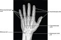 X-ray showing joints
 -
Hand bone anatomy


See also
- Dactylonomy
- Dermatoglyphics
- Finger-counting
- Finger tracking
- Handstand
- Hand strength
- Hand walking
- Human skeletal changes due to bipedalism
- Knuckle-walking
- Palmistry—fortune-telling based on lines in hand palms
- Manus (anatomy)
- Mudra—Hindu term for hand gestures
References
- ISBN 978-1-930665-67-5.)
{{cite book}}: CS1 maint: multiple names: authors list (link - ^ S2CID 4358448.)
{{cite journal}}: CS1 maint: multiple names: authors list (link - ^ ISBN 978-0-19-533316-9.
- ISBN 978-1-4939-3646-5.
- ISBN 9780199763108.
- ISBN 9780721669908.
- ^ ISBN 978-1-58890-007-4.
- ISBN 978-0-321-20413-4.
- ^ "Body Segment Data". ExRx.net.
- ISBN 978-1-85317-544-2.
- ^ Saladin, Kenneth S. (2007) Anatomy & Physiology: The Unity of Form and Function. New York, NY: McGraw-Hill.
- ^ ISBN 978-1-85317-544-2.
- ISBN 978-0-8036-1191-7.
- PMID 30969632. Retrieved November 28, 2020.
- ^ ISBN 978-0-19-517315-4.
- ISBN 978-1-58890-419-5.
- ISBN 978-0-19-517315-4.
- ^ "Web". Oxford English Dictionary (Online ed.). Oxford University Press. (Subscription or participating institution membership required.)
- ^ "web of fingers/ toes". Farlex Medical Dictionary. Farlex Partner Medical Dictionary. Retrieved 14 March 2016.
- PMID 29565244.
- ^ a b
Schmidt, Hans-Martin; Lanz, Ulrich (2003). Surgical Anatomy of the Hand. Thieme. p. 1. ISBN 978-1-58890-007-4.
- ^ .
- ^ Marzke, Mary. "Evolution of the hand and bipedality". Massey University, NZ. Retrieved September 21, 2017.
- ^ Flanagan, J Randall; Johansson, Roland S (2002). "Hand Movements" (PDF). Encyclopedia of the human brain. Elsevier Science.
- PMID 10637723.
- ISBN 978-84-693-4823-9. Retrieved 2023-03-25.
- PMID 19667206.
- ^ S2CID 19629241.
- PMID 18380869.)
- ^ Reardon, Sara (December 19, 2012). "Human hands evolved so we could punch each other". New Scientist. Retrieved September 21, 2017.
- .
- PMID 23255192.
External links
 Definitions from Wiktionary
 Media from Commons
 News from Wikinews
 Quotations from Wikiquote
 Texts from Wikisource
 Textbooks from Wikibooks
 Resources from Wikiversity
- Hand anatomy (eMedicine)
- Film Board of Canada documentary Faces of the Hand
- "The Common Hand" Archived 2016-07-13 at the National Geographic