Congenital diaphragmatic hernia
| Congenital diaphragmatic hernia | |
|---|---|
| Other names | CDH |
 | |
| Morgagni hernia seen on a chest radiograph. | |
| Specialty | Medical genetics, pediatrics |
Congenital diaphragmatic hernia (CDH) is a
CDH is a life-threatening pathology in infants and a major cause of death due to two complications:
Classification
Bochdalek hernia
The Bochdalek hernia, also known as a postero-lateral diaphragmatic hernia, is the most common manifestation of CDH, accounting for more than 95% of cases. In this instance the diaphragm abnormality is characterized by a hole in the postero-lateral corner of the diaphragm which allows passage of the abdominal viscera into the chest cavity. The majority of Bochdalek hernias (80–85%) occur on the left side of the diaphragm, a large proportion of the remaining cases occur on the right side. To date, it carries a high mortality[3] and is an active area of clinical research.[citation needed]
Morgagni hernia

This rare anterior defect of the diaphragm is variably referred to as a Morgagni, retrosternal, or parasternal hernia. Accounting for approximately 2% of all CDH cases, it is characterized by herniation through the
Diaphragm eventration
The diagnosis of congenital diaphragmatic eventration is used when there is abnormal displacement (i.e. elevation) of part or all of an otherwise intact diaphragm into the chest cavity. This rare type of CDH occurs because in the region of eventration the diaphragm is thinner, allowing the abdominal viscera to protrude upwards.[citation needed]
Pathophysiology
There are genetic causes of CDH[5] including aneuploidies, chromosome copy number variants, and single gene mutations. Research implicates a few gene mutations including LONP1[6] and MYRF.[7] It involves three major defects:[citation needed]
- A failure of the diaphragm to completely close during development
- chest
- Pulmonary hypoplasia
Diagnosis

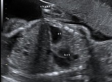
This condition can often be diagnosed before birth and
Survival rates for infants with this condition vary, but have generally been increasing through advances in neonatal medicine. Work has been done to correlate survival rates to ultrasound measurements of the lung volume as compared to the baby's head circumference. This figure known as the lung-to-head ratio (LHR). Still, LHR remains an inconsistent measure of survival. Outcomes of CDH are largely dependent on the severity of the defect and the appropriate timing of treatment.
A small percentage of cases go unrecognized into adulthood.[9]
Treatment
The first step in management is orogastric tube placement and securing the airway (intubation). Ideally, the baby will never take a breath, to avoid air going into the intestines and compressing the lungs and heart. The baby will then be immediately placed on a ventilator. Extracorporeal membrane oxygenation (ECMO) has been used as part of the treatment strategy at some hospitals.[10][11] ECMO acts as a heart-lung bypass.
Diaphragm eventration is typically repaired thoracoscopically, by a technique called plication of the diaphragm.[12] Plication basically involves a folding of the eventrated diaphragm which is then sutured in order to “take up the slack” of the excess diaphragm tissue.[citation needed]
Prognosis
Congenital diaphragmatic hernia has a mortality rate of 40–62%,[13] with outcomes being more favorable in the absence of other congenital abnormalities. Individual rates vary greatly dependent upon multiple factors: size of hernia, organs involved, additional birth defects and/or genetic problems, amount of lung growth, age and size at birth, type of treatments, timing of treatments, complications (such as infections) and lack of lung function.[citation needed]
See also
References
- PMID 19154527.
- PMID 17848243.
- PMID 25087794.
- PMID 19527083.
- PMID 25447988.
- PMID 34547244.
- S2CID 54480742.
- ^ "Deadly hernia corrected in womb – Surgeons have developed an operation to repair a potentially fatal abnormality in babies before they are born". BBC News. 2004-07-26. Retrieved 2006-07-14. – report of new operation, pioneered at London's King's College Hospital which reduced death rates in the most at risk by 50%
- S2CID 30028643.
- PMID 17706494.
- S2CID 15451125.
- PMID 16291157.
- ^ Pediatric Congenital Diaphragmatic Hernia at eMedicine
External links
- Congenital Diaphragmatic Hernia Study Group under University of Texas Health Science Center at Houston
- DHREAMS (Diaphragmatic Hernia Research & Exploration; Advancing Molecular Science) Project coordinated at Columbia University Medical Center
- CDH International – A global initiative to stop Congenital Diaphragmatic Hernia
