Progesterone receptor
Ensembl | |||||||||
|---|---|---|---|---|---|---|---|---|---|
| UniProt | |||||||||
| RefSeq (mRNA) | |||||||||
| RefSeq (protein) | |||||||||
| Location (UCSC) | Chr 11: 101.03 – 101.13 Mb | Chr 9: 8.9 – 8.97 Mb | |||||||
| PubMed search | [3] | [4] | |||||||
| View/Edit Human | View/Edit Mouse |
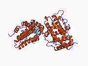
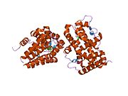
The progesterone receptor (PR), also known as NR3C3 or nuclear receptor subfamily 3, group C, member 3, is a protein found inside cells. It is activated by the steroid hormone progesterone.
In humans, PR is encoded by a single PGR gene residing on chromosome 11q22,[5][6][7] it has two isoforms, PR-A and PR-B, that differ in their molecular weight.[8][9][10] The PR-B is the positive regulator of the effects of progesterone, while PR-A serve to antagonize the effects of PR-B.[11]
Mechanism
After progesterone binds to the receptor, restructuring with dimerization follows and the complex enters the nucleus and binds to DNA. There transcription takes place, resulting in formation of messenger RNA that is translated by ribosomes to produce specific proteins.
Structure
| Progesterone receptor, N-terminal | |||||||||
|---|---|---|---|---|---|---|---|---|---|
| Identifiers | |||||||||
| Symbol | Progest_rcpt_N | ||||||||
| Pfam | PF02161 | ||||||||
| InterPro | IPR000128 | ||||||||
| |||||||||
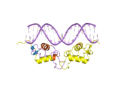
In common with other steroid receptors, the progesterone receptor has a
Isoforms
As demonstrated in progesterone receptor-deficient mice, the physiological effects of progesterone depend completely on the presence of the human progesterone receptor (hPR), a member of the steroid-receptor superfamily of nuclear receptors. The single-copy human (hPR) gene uses separate promoters and translational start sites to produce two isoforms, hPR-A and -B, which are identical except for an additional 165 amino acids present only in the N terminus of hPR-B.[12] Although hPR-B shares many important structural domains with hPR-A, they are in fact two functionally distinct transcription factors, mediating their own response genes and physiological effects with little overlap. Selective ablation of PR-A in a mouse model, resulting in exclusive production of PR-B, unexpectedly revealed that PR-B contributes to, rather than inhibits, epithelial cell proliferation both in response to estrogen alone and in the presence of progesterone and estrogen. These results suggest that in the uterus, the PR-A isoform is necessary to oppose estrogen-induced proliferation as well as PR-B-dependent proliferation.
Functional polymorphisms
Six variable sites, including four polymorphisms and five common haplotypes have been identified in the human PR gene .[13] One promoter region polymorphism, +331G/A, creates a unique transcription start site. Biochemical assays showed that the +331G/A polymorphism increases transcription of the PR gene, favoring production of hPR-B in an Ishikawa endometrial cancer cell line.[14]
Several studies have now shown no association between progesterone receptor gene +331G/A polymorphisms and breast or endometrial cancers.[15][16] However, these follow-up studies lacked the sample size and statistical power to make any definitive conclusions, due to the rarity of the +331A SNP. It is currently unknown which if any polymorphisms in this receptor are of significance to cancer. A study of 21 non-European populations identified two markers within the PROGINS haplotype of the PR gene as positively correlated with ovarian and breast cancer.[17]
Animal studies
Development
Knockout[
Behavior]
During rodent perinatal life, progesterone receptor (PR) is known to be transiently expressed in both the ventral tegmental area (VTA) and the medial prefrontal cortex (mPFC) of the mesocortical dopaminergic pathway. PR activity during this time period impacts the development of dopaminergic innervation of the mPFC from the VTA. If PR activity is altered, a change in dopaminergic innervation of the mPFC is seen and tyrosine hydroxylase (TH), the rate-limiting enzyme for dopamine synthesis, in the VTA will also be impacted. TH expression in this area is an indicator of dopaminergic activity, which is believed to be involved in normal and critical development of complex cognitive behaviors that are mediated by the mesocortical dopaminergic pathway, such as working memory, attention, behavioral inhibition, and cognitive flexibility.[21]
Research has shown that when a PR antagonist, such as RU 486, is administered to rats during the neonatal period, decreased tyrosine hydroxylase immunoreactive (TH-ir) cells density, a strong co-expresser with PR-immunoreactivity (PR-ir), is seen in the mPFC of juvenile rodents. Later on, in adulthood, decreased levels of TH-ir in the VTA are also shown. This alteration in TH-ir fiber expression, an indicator of altered dopaminergic activity resulting from neonatal PR antagonist administration, has been shown to impair later performance on tasks that measure behavioral inhibition and impulsivity, as well as cognitive flexibility in adulthood. Similar cognitive flexibility impairments were also seen in PR knockout mice as a result of reduced dopaminergic activity in the VTA.[21]
Conversely, when a PR agonist, such as 17α-hydroxyprogesterone caproate, is administered to rodents during perinatal life, as the mesocortical dopaminergic pathway is developing, dopaminergic innervation of the mPFC increases. As a result, TH-ir fiber density also increases. Interestingly, this increase in TH-ir fibers and dopaminergic activity is also linked to impaired cognitive flexibility with increased perseveration later on in life.[22]
In combination, these findings suggest that PR expression during early development impact later cognitive functioning in rodents. Furthermore, it appears as though abnormal levels of PR activity during this critical period of mesocortical dopaminergic pathway development may have profound effects on specific behavioral neural circuits involved in the formation of later complex cognitive behavior.[21][22]
Ligands
Agonists
- Endogenous progestogens (e.g., progesterone)
- Synthetic progestogens (e.g., norethisterone, levonorgestrel, medroxyprogesterone acetate, megestrol acetate, dydrogesterone, drospirenone)
Mixed
Antagonists
- Antiprogestogens (e.g., mifepristone, aglepristone, onapristone, lonaprisan, lilopristone, toripristone)[23]
Interactions
Progesterone receptor has been shown to
See also
References
- ^ a b c GRCh38: Ensembl release 89: ENSG00000082175 – Ensembl, May 2017
- ^ a b c GRCm38: Ensembl release 89: ENSMUSG00000031870 – Ensembl, May 2017
- ^ "Human PubMed Reference:". National Center for Biotechnology Information, U.S. National Library of Medicine.
- ^ "Mouse PubMed Reference:". National Center for Biotechnology Information, U.S. National Library of Medicine.
- PMID 3551956.
- PMID 3472240.
- ^ ensembl.org, Gene: ESR1 (ENSG00000091831)
- PMID 15970482.
- ISBN 978-0-683-30379-7.
- ISBN 978-0-7817-4795-0.
- ISBN 978-1-4614-6837-0.
- PMID 2328727.
- PMID 15718480.
- PMID 12218173.
- S2CID 46282046.
- PMID 16835347.
- S2CID 205302494.
- PMID 22844349.
- PMID 26266959.
- PMID 23705924.
- ^ PMID 26065828.
- ^ PMID 26556535.
- ^ PMID 24291072.
- PMID 12672823.
- PMID 10757795.
- PMID 9891052.
Further reading
- Butnor KJ, Burchette JL, Robboy SJ (July 1999). "Progesterone receptor activity in leiomyomatosis peritonealis disseminata". International Journal of Gynecological Pathology. 18 (3): 259–64. PMID 12090595.
- Leonhardt SA, Boonyaratanakornkit V, Edwards DP (November 2003). "Progesterone receptor transcription and non-transcription signaling mechanisms". Steroids. 68 (10–13): 761–70. S2CID 7533810.
- Conneely OM, Mulac-Jericevic B, Lydon JP (November 2003). "Progesterone-dependent regulation of female reproductive activity by two distinct progesterone receptor isoforms". Steroids. 68 (10–13): 771–8. S2CID 13600266.
- Bagchi MK, Tsai SY, Tsai MJ, O'Malley BW (April 1992). "Ligand and DNA-dependent phosphorylation of human progesterone receptor in vitro". Proceedings of the National Academy of Sciences of the United States of America. 89 (7): 2664–8. PMID 1557371.
- Kastner P, Krust A, Turcotte B, Stropp U, Tora L, Gronemeyer H, Chambon P (May 1990). "Two distinct estrogen-regulated promoters generate transcripts encoding the two functionally different human progesterone receptor forms A and B". The EMBO Journal. 9 (5): 1603–14. PMID 2328727.
- Guiochon-Mantel A, Loosfelt H, Lescop P, Sar S, Atger M, Perrot-Applanat M, Milgrom E (June 1989). "Mechanisms of nuclear localization of the progesterone receptor: evidence for interaction between monomers". Cell. 57 (7): 1147–54. PMID 2736623.
- Fernandez MD, Carter GD, Palmer TN (January 1983). "The interaction of canrenone with oestrogen and progesterone receptors in human uterine cytosol". British Journal of Clinical Pharmacology. 15 (1): 95–101. PMID 6849751.
- Oñate SA, Tsai SY, Tsai MJ, O'Malley BW (November 1995). "Sequence and characterization of a coactivator for the steroid hormone receptor superfamily". Science. 270 (5240): 1354–7. S2CID 28749162.
- Zhang Y, Beck CA, Poletti A, Edwards DP, Weigel NL (December 1994). "Identification of phosphorylation sites unique to the B form of human progesterone receptor. In vitro phosphorylation by casein kinase II". The Journal of Biological Chemistry. 269 (49): 31034–40. PMID 7983041.
- Mansour I, Reznikoff-Etievant MF, Netter A (August 1994). "No evidence for the expression of the progesterone receptor on peripheral blood lymphocytes during pregnancy". Human Reproduction. 9 (8): 1546–9. PMID 7989520.
- Kalkhoven E, Wissink S, van der Saag PT, van der Burg B (March 1996). "Negative interaction between the RelA(p65) subunit of NF-kappaB and the progesterone receptor". The Journal of Biological Chemistry. 271 (11): 6217–24. PMID 8626413.
- Wang JD, Zhu JB, Fu Y, Shi WL, Qiao GM, Wang YQ, Chen J, Zhu PD (February 1996). "Progesterone receptor immunoreactivity at the maternofetal interface of first trimester pregnancy: a study of the trophoblast population". Human Reproduction. 11 (2): 413–9. PMID 8671234.
- Thénot S, Henriquet C, Rochefort H, Cavaillès V (May 1997). "Differential interaction of nuclear receptors with the putative human transcriptional coactivator hTIF1". The Journal of Biological Chemistry. 272 (18): 12062–8. PMID 9115274.
- Jenster G, Spencer TE, Burcin MM, Tsai SY, Tsai MJ, O'Malley BW (July 1997). "Steroid receptor induction of gene transcription: a two-step model". Proceedings of the National Academy of Sciences of the United States of America. 94 (15): 7879–84. PMID 9223281.
- Shanker YG, Sharma SC, Rao AJ (September 1997). "Expression of progesterone receptor mRNA in the first trimester human placenta". Biochemistry and Molecular Biology International. 42 (6): 1235–40. S2CID 24959703.
- Richer JK, Lange CA, Wierman AM, Brooks KM, Tung L, Takimoto GS, Horwitz KB (April 1998). "Progesterone receptor variants found in breast cells repress transcription by wild-type receptors". Breast Cancer Research and Treatment. 48 (3): 231–41. S2CID 27266907.
- Williams SP, Sigler PB (May 1998). "Atomic structure of progesterone complexed with its receptor". Nature. 393 (6683): 392–6. S2CID 4424486.
- Boonyaratanakornkit V, Melvin V, Prendergast P, Altmann M, Ronfani L, Bianchi ME, Taraseviciene L, Nordeen SK, Allegretto EA, Edwards DP (August 1998). "High-mobility group chromatin proteins 1 and 2 functionally interact with steroid hormone receptors to enhance their DNA binding in vitro and transcriptional activity in mammalian cells". Molecular and Cellular Biology. 18 (8): 4471–87. PMID 9671457.
- Nawaz Z, Lonard DM, Smith CL, Lev-Lehman E, Tsai SY, Tsai MJ, O'Malley BW (February 1999). "The Angelman syndrome-associated protein, E6-AP, is a coactivator for the nuclear hormone receptor superfamily". Molecular and Cellular Biology. 19 (2): 1182–9. PMID 9891052.
External links
- Progesterone+Receptors at the U.S. National Library of Medicine Medical Subject Headings (MeSH)


