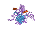
CCL4
Chemokine (C-C motif) ligands 4 (also CCL4) previously known as macrophage inflammatory protein (MIP-1β), is a protein which in humans is encoded by the CCL4 gene.[2] CCL4 belongs to a cluster of genes located on 17q11-q21 of the chromosomal region.[3] Identification and localization of the gene on the chromosome 17 was in 1990 although the discovery of MIP-1 was initiated in 1988 with the purification of a protein doublet corresponding to inflammatory activity from supernatant of endotoxin-stimulated murine macrophages. At that time, it was also named as "macrophage inflammatory protein-1" (MIP-1) due to its inflammatory properties.[4]
CCL4 is a small cytokine that belongs to the CC chemokine subfamily. CCL4 is being secreted under mitogenic signals and antigens and hereby acts as a chemoattractant for natural killer cells, monocytes and various other immune cells in the site of inflamed or damaged tissue.[5]
Genomics
In the human genome, CCL4 and many other CC chemokines is encoded by a single gene on chromosome 17 (17q11-q21). The CCL4 gene consists of three exons and two introns which are separated by 14 kb and are organized in a head to head fashion. MIP-1 genes have 3 untranslated gene regions containing a polyadenylation site (AATAAA) and several AT-rich sequences.[6] The CCL4 protein precursor consist of 92 amino acids. In turn, the mature CCL4 protein is 92 amino acids long. The CCL4 predicted Mr weight is 7814.8 Da with no apparent N-linked glycosylation site as in other of the MIP-1 proteins.[7][8][9]

Molecular structure
CCL4 is a polypeptide chain with a
Monomeric subunits in their secondary structure composed by a triple-stranded antiparallel sheet form in a Greek key structure on top of which lies an α-helix. NH2-terminus is arranged as a long loop followed by a four-residue helical turn. The overall form of homodimer is globular elongated and cylindrical with sizes: 56 Å × 30 Å × 26 Å in contrast of monomer structure which is similar to IL-8.[10][11]
CCL4 as well as other MIP-1s whether human or mouse have a high tendency to self-aggregation. Aggregation as a reversible and dynamic process depends largely on the concentration of chemokine.[12]
The distinction between the CC chemokine families, MIP-1α and MIP-1β, was initially based on whether the first two cysteine residues are separated by one residue (α) or are adjacent (β).[10] Final form of tertiary structure structure of MIP-1 has been defined by heteronuclear magnetic resonance (NMR) analysis.
Concentration of this chemokine has been shown to be inversely related with MicroRNA-125b. Concentration of CCL4 within the body increases with age, which may cause chronic inflammation and liver damage.[13][14]
Function
CCL4 as a chemokine which is produced during inflammation, damage or other important dynamic processes as an angiogenesis to attract immune cells as leukocytes transgress the vascular endothelium and migrate into peripheral tissues.
Production of CCL4
CCL4 is produced by: monocytes, B cells, T cells, NK cells, dendritic cells, neutrophils, fibroblasts, endothelial cells such as vascular smooth muscle cells, brain microvessel endothelial cells, fetal microglia and epithelial cells.
- Monocytes produce high amounts of CCL4 when they are stimulated with LPS or IL-7 and production is suppressed by IL-4.[15]
- T cells and B cells secrete CCL4 response to Ag receptor (BCR) triggering.[16]
- NK cells produce CCL4 in response to stimulation with IL-2, physiological activation signals such as lysis. NK cells can be important source of CC chemokines and may suppress HIV infection by inhibition replication of HIV-1 virus by interfering with the ability to utilize CCR5 as a coreceptor for entry in CD4(+) cells.[17]
- Dendritic cells secrete and respond to chemokines after stimulation to LPS, TNFα, or CD40 ligand. In maturing of DC production of chemokines have different impact on chemokine receptor function: CCR1 and CCR5 were down-regulated whereas CCR7 were stimulated increased in maturing DC. Production of chemokines provides DC with the capacity to self-regulate their migratory behavior as well as to recruit other cells.[18]
- Neutrophils produce CCL4 in the presence of IFN-gamma and its inhibition by IL-10.[19]
- Endothelial cells release CCL4 following stimulation with LPS, TNFα, IFN-, or IL-1.[20]
CCL4 is a major HIV-suppressive factor produced by CD8+ T cells.[21]
CCL4 is produced by: neutrophils, monocytes, B cells, T cells, fibroblasts, endothelial cells, and epithelial cells.[13]
Interactions
CCL4 has been shown to
CCL4 binds to G protein-Coupled Receptors
See also
References
- ^ "Human PubMed Reference:". National Center for Biotechnology Information, U.S. National Library of Medicine.
- PMID 1972563.
- PMID 30123055.
- PMID 3279154.
- PMID 12401480.
- PMID 12401480.
- PMID 1296815.
- PMID 2462251.
- PMID 9092555.
- ^ PMID 8134838.
- PMID 10347159.
- ^ Nibbs RJ, Graham GJ, Pragnell IB (1998). "Macrophage Inflammatory Protein 1-α". Cytokines. Elsevier. pp. 467–488.
- ^ PMID 25620312.
- PMID 3887178.
- PMID 1596574.
- PMID 11001880.
- PMID 9649576.
- S2CID 13766954.
- PMID 11083814.
- PMID 10888363.
- S2CID 84062618.
- PMID 11470920.
- PMID 11278300.
Further reading
- Menten P, Wuyts A, Van Damme J (December 2002). "Macrophage inflammatory protein-1". Cytokine & Growth Factor Reviews. 13 (6): 455–481. PMID 12401480.
- Muthumani K, Desai BM, Hwang DS, Choo AY, Laddy DJ, Thieu KP, et al. (April 2004). "HIV-1 Vpr and anti-inflammatory activity". DNA and Cell Biology. 23 (4): 239–247. PMID 15142381.
- Conti L, Fantuzzi L, Del Cornò M, Belardelli F, Gessani S (2005). "Immunomodulatory effects of the HIV-1 gp120 protein on antigen presenting cells: implications for AIDS pathogenesis". Immunobiology. 209 (1–2): 99–115. PMID 15481145.
- Joseph AM, Kumar M, Mitra D (January 2005). "Nef: "necessary and enforcing factor" in HIV infection". Current HIV Research. 3 (1): 87–94. PMID 15638726.
- Zhao RY, Elder RT (March 2005). "Viral infections and cell cycle G2/M regulation". Cell Research. 15 (3): 143–149. PMID 15780175.
- Zhao RY, Bukrinsky M, Elder RT (April 2005). "HIV-1 viral protein R (Vpr) & host cellular responses". The Indian Journal of Medical Research. 121 (4): 270–286. PMID 15817944.
- Li L, Li HS, Pauza CD, Bukrinsky M, Zhao RY (2006). "Roles of HIV-1 auxiliary proteins in viral pathogenesis and host-pathogen interactions". Cell Research. 15 (11–12): 923–934. PMID 16354571.
- King JE, Eugenin EA, Buckner CM, Berman JW (April 2006). "HIV tat and neurotoxicity". Microbes and Infection. 8 (5): 1347–1357. PMID 16697675.
- Napolitano M, Modi WS, Cevario SJ, Gnarra JR, Seuanez HN, Leonard WJ (September 1991). "The gene encoding the Act-2 cytokine. Genomic structure, HTLV-I/Tax responsiveness of 5' upstream sequences, and chromosomal localization". The Journal of Biological Chemistry. 266 (26): 17531–17536. PMID 1894635.
- Irving SG, Zipfel PF, Balke J, McBride OW, Morton CC, Burd PR, et al. (June 1990). "Two inflammatory mediator cytokine genes are closely linked and variably amplified on chromosome 17q". Nucleic Acids Research. 18 (11): 3261–3270. PMID 1972563.
- Baixeras E, Roman-Roman S, Jitsukawa S, Genevee C, Mechiche S, Viegas-Pequignot E, et al. (November 1990). "Cloning and expression of a lymphocyte activation gene (LAG-1)". Molecular Immunology. 27 (11): 1091–1102. PMID 2247088.
- Lipes MA, Napolitano M, Jeang KT, Chang NT, Leonard WJ (December 1988). "Identification, cloning, and characterization of an immune activation gene". Proceedings of the National Academy of Sciences of the United States of America. 85 (24): 9704–9708. PMID 2462251.
- Brown KD, Zurawski SM, Mosmann TR, Zurawski G (January 1989). "A family of small inducible proteins secreted by leukocytes are members of a new superfamily that includes leukocyte and fibroblast-derived inflammatory agents, growth factors, and indicators of various activation processes". Journal of Immunology. 142 (2): 679–687. PMID 2521353.
- Zipfel PF, Balke J, Irving SG, Kelly K, Siebenlist U (March 1989). "Mitogenic activation of human T cells induces two closely related genes which share structural similarities with a new family of secreted factors". Journal of Immunology. 142 (5): 1582–1590. PMID 2521882.
- Chang HC, Reinherz EL (June 1989). "Isolation and characterization of a cDNA encoding a putative cytokine which is induced by stimulation via the CD2 structure on human T lymphocytes". European Journal of Immunology. 19 (6): 1045–1051. S2CID 35270650.
- Miller MD, Hata S, De Waal Malefyt R, Krangel MS (November 1989). "A novel polypeptide secreted by activated human T lymphocytes". Journal of Immunology. 143 (9): 2907–2916. PMID 2809212.
- Adams MD, Kerlavage AR, Fleischmann RD, Fuldner RA, Bult CJ, Lee NH, et al. (September 1995). "Initial assessment of human gene diversity and expression patterns based upon 83 million nucleotides of cDNA sequence". Nature. 377 (6547 Suppl): 3–174. PMID 7566098.
- Post TW, Bozic CR, Rothenberg ME, Luster AD, Gerard N, Gerard C (December 1995). "Molecular characterization of two murine eosinophil beta chemokine receptors". Journal of Immunology. 155 (11): 5299–5305. PMID 7594543.
- Combadiere C, Ahuja SK, Murphy PM (July 1995). "Cloning and functional expression of a human eosinophil CC chemokine receptor". The Journal of Biological Chemistry. 270 (28): 16491–16494. PMID 7622448.
- Paolini JF, Willard D, Consler T, Luther M, Krangel MS (September 1994). "The chemokines IL-8, monocyte chemoattractant protein-1, and I-309 are monomers at physiologically relevant concentrations". Journal of Immunology. 153 (6): 2704–2717. PMID 8077676.
External links
- Human CCL4 genome location and CCL4 gene details page in the UCSC Genome Browser.


