
Ear
| Ear | |
|---|---|
.jpg) The outer portion of the human ear "Ear" pronounced (Received Pronunciation) | |
| Details | |
| System | Auditory system |
| Identifiers | |
| Latin | auris |
| MeSH | D004423 |
| NeuroLex ID | birnlex_1062 |
| TA98 | A01.1.00.005 A15.3.00.001 |
| TA2 | 6861 |
| FMA | 52780 |
| Anatomical terminology | |
 |
| This article is one of a series documenting the anatomy of the |
| Human ear |
|---|
An ear is the
The ear develops from the first pharyngeal pouch and six small swellings that develop in the early embryo called otic placodes, which are derived from ectoderm.
The ear may be affected by disease, including infection and traumatic damage. Diseases of the ear may lead to
The ear has been adorned by earrings and other jewelry in numerous cultures for thousands of years, and has been subjected to surgical and cosmetic alterations.
Structure
The human ear consists of three parts—the
Outer ear
The outer ear is the external portion of the ear and includes the fleshy visible auricle, the ear canal, and the outer layer of the eardrum (also called the tympanic membrane).[6][7]
The auricle consists of the curving outer rim called the
Two sets of muscles are associated with the outer ear: the
The auricle consists of a single piece of
Middle ear

The middle ear lies between the outer ear and the inner ear. It consists of an air-filled cavity called the
The three ossicles transmit sound from the outer ear to the inner ear. The malleus receives vibrations from sound pressure on the eardrum, where it is connected at its longest part (the manubrium or handle) by a ligament. It transmits vibrations to the incus, which in turn transmits the vibrations to the small stapes bone. The wide base of the stapes rests on the oval window. As the stapes vibrates, vibrations are transmitted through the oval window, causing movement of fluid within the cochlea.[7]
The round window allows for the fluid within the inner ear to move. As the stapes pushes the
Inner ear

The inner ear sits within the temporal bone in a complex cavity called the
The bony labyrinth refers to the bony compartment which contains the membranous labyrinth, contained within the temporal bone. The inner ear structurally begins at the oval window, which receives vibrations from the incus of the middle ear. Vibrations are transmitted into the inner ear into a fluid called endolymph, which fills the membranous labyrinth. The endolymph is situated in two vestibules, the utricle and saccule, and eventually transmits to the cochlea, a spiral-shaped structure. The cochlea consists of three fluid-filled spaces: the vestibular duct, the cochlear duct, and the tympanic duct.[7] Hair cells responsible for transduction—changing mechanical changes into electrical stimuli are present in the organ of Corti in the cochlea.[12]
Blood supply
The blood supply of the ear differs according to each part of the ear.
The outer ear is supplied by a number of arteries. The posterior auricular artery provides the majority of the blood supply. The anterior auricular arteries provide some supply to the outer rim of the ear and scalp behind it. The posterior auricular artery is a direct branch of the external carotid artery, and the anterior auricular arteries are branches from the superficial temporal artery. The occipital artery also plays a role.[12]
The middle ear is supplied by the mastoid branch of either the occipital or posterior auricular arteries and the deep auricular artery, a branch of the maxillary artery. Other arteries which are present but play a smaller role include branches of the middle meningeal artery, ascending pharyngeal artery, internal carotid artery, and the artery of the pterygoid canal.[12]
The inner ear is supplied by the anterior tympanic branch of the maxillary artery; the stylomastoid branch of the posterior auricular artery; the petrosal branch of middle meningeal artery; and the labyrinthine artery, arising from either the anterior inferior cerebellar artery or the basilar artery.[12]
Function
Hearing
Sound that travels through the outer ear impacts on the eardrum, and causes it to vibrate. The three ossicles bones transmit this sound to a second window (the
The inner ear houses the apparatus necessary to
The human ear can generally hear sounds with frequencies between 20
Balance
Providing balance, when moving or stationary, is also a central function of the ear. The ear facilitates two types of balance: static balance, which allows a person to feel the effects of gravity, and dynamic balance, which allows a person to sense acceleration.
Static balance is provided by two ventricles, the utricle and the saccule. Cells lining the walls of these ventricles contain fine filaments, and the cells are covered with a fine gelatinous layer. Each cell has 50–70 small filaments, and one large filament, the
Dynamic balance is provided through the three semicircular canals. These three canals are orthogonal (at right angles) to each other. At the end of each canal is a slight enlargement, known as the
Development
During
Inner ear
.png)
After implantation, around the second to third week the
Around the 33rd day of development, the vesicles begin to differentiate. Closer to the back of the embryo, they form what will become the utricle and semicircular canals. Closer to the front of the embryo, the vesicles differentiate into a rudimentary saccule, which will eventually become the saccule and cochlea. Part of the saccule will eventually give rise and connect to the cochlear duct. This duct appears approximately during the sixth week and connects to the saccule through the ductus reuniens.[18]
As the cochlear duct's mesenchyme begins to differentiate, three cavities are formed: the
Parts of the otic vesicle in turn form the vestibulocochlear nerve.[22] These form bipolar neurons, which supply sensation to parts of the inner ear (namely the sensory parts of the semicircular canals, macular of the utricle and saccule, and organ of Corti). The nerve begins to form around the 28th day.[20]
- Molecular regulation
Most of the genes responsible for the
Middle ear
The middle ear and its components develop from the first and second
Outer ear

Unlike structures of the inner and middle ear, which develop from pharyngeal pouches, the ear canal originates from the dorsal portion of the first
Clinical significance
Hearing loss


Hearing loss may be either partial or total. This may be a result of injury or damage,
Causes of conductive hearing loss include an ear canal blocked by ear wax, ossicles that are fixed together or absent, or holes in the eardrum. Conductive hearing loss may also result from middle ear inflammation causing fluid build-up in the normally air-filled space, such as by otitis media. Tympanoplasty is the general name of the operation to repair the middle ear's eardrum and ossicles. Grafts from muscle fascia are ordinarily used to rebuild an intact eardrum. Sometimes artificial ear bones are placed to substitute for damaged ones, or a disrupted ossicular chain is rebuilt in order to conduct sound effectively.
Hearing aids or cochlear implants may be used if the hearing loss is severe or prolonged. Hearing aids work by amplifying the sound of the local environment and are best suited to conductive hearing loss.[24] Cochlear implants transmit the sound that is heard as if it were a nervous signal, bypassing the cochlea. Active middle ear implants send sound vibrations to the ossicles in the middle ear, bypassing any non-functioning parts of the outer and middle ear.
Congenital abnormalities
Anomalies and malformations of the auricle are common. These anomalies include chromosome syndromes such as
Approximately one out of one thousand children suffer some type of congenital deafness related to the development of the inner ear.
Vertigo
Vertigo refers to the inappropriate perception of motion. This is due to dysfunction of the vestibular system. One common type of vertigo is benign paroxysmal positional vertigo, when an otolith is displaced from the ventricles to the semicircular canal. The displaced otolith rests on the cupola, causing a sensation of movement when there is none. Ménière's disease, labyrinthitis, strokes, and other infective and congenital diseases may also result in the perception of vertigo.[30]
Injury
- Outer ear
Injuries to the external ear occur fairly frequently, and can leave minor to major deformity. Injuries include:
- Middle ear
The ear drum may become perforated in the event of a large sound or explosion, when diving or flying (called
- Inner ear
There are two principal damage mechanisms to the inner ear in industrialised society, and both injure hair cells. The first is exposure to elevated sound levels (noise trauma), and the second is exposure to drugs and other substances (
Tinnitus
Tinnitus is the hearing of sound when no external sound is present.[43] While often described as a ringing, it may also sound like a clicking, hiss or roaring.[44] Rarely, unclear voices or music are heard.[45] The sound may be soft or loud, low pitched or high pitched and appear to be coming from one ear or both.[44] Most of the time, it comes on gradually.[45] In some people, the sound causes depression, anxiety, or concentration difficulties.[44]
Tinnitus is not a disease but a symptom that can result from a number of underlying causes. One of the most common causes is
Society and culture

The ears have been ornamented with jewelry for thousands of years, traditionally by
Injury to the ears has been present since Roman times as a method of reprimand or punishment – "In Roman times, when a dispute arose that could not be settled amicably, the injured party cited the name of the person thought to be responsible before the Praetor; if the offender did not appear within the specified time limit, the complainant summoned witnesses to make statements. If they refused, as often happened, the injured party was allowed to drag them by the ear and to pinch them hard if they resisted. Hence the French expression "se faire tirer l’oreille", of which the literal meaning is "to have one's ear pulled" and the figurative meaning "to take a lot of persuading". We use the expression "to tweak (or pull) someone's ears" to mean "inflict a punishment"."[31]
The auricles have an effect on facial appearance. In Western societies, protruding ears (present in about 5% of ethnic

Georg von Békésy was a Hungarian biophysicist born in Budapest, Hungary. In 1961, he was awarded the Nobel Prize in Physiology or Medicine for his research on the function of the cochlea in the mammalian hearing organ.[66]
The Vacanti mouse was a laboratory mouse that had what looked like a human ear grown on its back. The "ear" was actually an ear-shaped cartilage structure grown by seeding cow cartilage cells into a biodegradable ear-shaped mold and then implanted under the skin of the mouse; then the cartilage naturally grew by itself.[67] It was developed as an alternative to ear repair or grafting procedures and the results met with much publicity and controversy in 1997.[68][69]
Other animals

The pinna helps direct sound through the ear canal to the eardrum. The complex geometry of ridges on the inner surface of some mammalian ears helps to sharply focus sounds produced by prey, using echolocation signals. These ridges can be regarded as the acoustic equivalent of a
Some large
In some animals with mobile pinnae (like the horse), each pinna can be aimed independently to better receive the sound. For these animals, the pinnae help localise the direction of the sound source.
-
 African bush elephant
Loxodonta africana -
 Fennec fox (desert regions)
Vulpes zerda -
 Arctic fox
Vulpes lagopus -
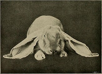


Illustration by
Charles Darwin, 1868
The ear, with its blood vessels close to the surface, is an essential thermoregulator in some land mammals, including the elephant, the fox, and the rabbit.[75] There are five types of ear carriage in domestic rabbits, some of which have been bred for exaggerated ear length[76]—a potential health risk that is controlled in some countries.[77] Abnormalities in the skull of a half-lop rabbit were studied by Charles Darwin in 1868. In marine mammals, earless seals are one of three groups of Pinnipedia.
Invertebrates
Only vertebrate animals have ears, though many invertebrates detect sound using other kinds of sense organs. In insects, tympanal organs are used to hear distant sounds. They are located either on the head or elsewhere, depending on the insect family.[78] The tympanal organs of some insects are extremely sensitive, offering acute hearing beyond that of most other animals. The female cricket fly Ormia ochracea has tympanal organs on each side of her abdomen. They are connected by a thin bridge of exoskeleton and they function like a tiny pair of eardrums, but, because they are linked, they provide acute directional information. The fly uses her "ears" to detect the call of her host, a male cricket. Depending on where the song of the cricket is coming from, the fly's hearing organs will reverberate at slightly different frequencies. This difference may be as little as 50 billionths of a second, but it is enough to allow the fly to home in directly on a singing male cricket and parasitise it.[79]
Simpler structures allow other arthropods to detect near-field sounds. Spiders and cockroaches, for example, have hairs on their legs, which are used for detecting sound. Caterpillars may also have hairs on their body that perceive vibrations[80] and allow them to respond to sound.
See also
References
- ^ "Ear". Oxford Dictionary. Archived from the original on 18 July 2012. Retrieved 25 February 2016.
- ^ Shmerling, Robert H. (17 May 2017). "3 reasons to leave earwax alone". Harvard Health. Retrieved 11 July 2023.
- ^ "Why Do I Have So Much Earwax?". Keck Medicine of USC. 29 September 2022. Retrieved 11 July 2023.
- ^ "Earwax". HealthLink BC. 1 July 2021. Retrieved 11 July 2023.
- ^ "Earwax". MyHealth.Alberta.ca. 11 July 2023. Retrieved 11 July 2023.
- ^ ISBN 978-0-443-06684-9. Alt URL
- ^ ISBN 978-0-8089-2306-0.
- ISBN 978-1-4511-8447-1.
- ISBN 0-316-32268-7(P)
- ISBN 978-0-87893-697-7.
- ISBN 978-0-8089-2306-0.)
{{cite book}}: CS1 maint: multiple names: authors list (link - ^ ISBN 978-0-443-06684-9.
- ^ ISBN 978-0-7216-0240-0.)
{{cite book}}: CS1 maint: multiple names: authors list (link - ^ Greinwald, John H. Jr MD; Hartnick, Christopher J. MD The Evaluation of Children With Sensorineural Hearing Loss. Archives of Otolaryngology – Head & Neck Surgery. 128(1):84–87, January 2002
- ^ "Definition of "ultrasound" | Collins English Dictionary". www.collinsdictionary.com. Retrieved 20 March 2016.
- ISBN 978-0-7216-0240-0.)
{{cite book}}: CS1 maint: multiple names: authors list (link - ^ a b Moore, Keith L. (2009). Fundamentos de Anatomía con Orientación Clínica. pp. 1021–1035.
- ^ a b c d e f g h i Sadler, T.W. (2010). Embriología Médica. pp. 321–327.
- ^ a b c d e f Moore, Keith L. (2008). Embriología Clínica. pp. 477–482.
- ^ ISBN 978-0-443-06684-9.
- ^ a b c d e UNSW Embryology. Hearing-Inner Ear Development. Archived from the original on 30 September 2012. Retrieved 20 April 2013.
- ^ a b Drake, Richard L.; Wayne, A.; Mitchell, Adam (2010). GRAY Anatomía para estudiantes. pp. 854–871.
- PMID 20637105.
- ^ "Hearing Aids". National institute of deafness and other communication disorders. Retrieved 20 March 2016.
- ^ a b c Kliegman; Behrman; Jenson (2007). "367". Nelson Textbook of Pedriatics.
- ^ Lam SM. Edward Talbot Ely: father of aesthetic otoplasty. [Biography. Historical Article. Journal Article] Archives of Facial Plastic Surgery. 6(1):64, 2004 Jan–Feb.
- ^ Siegert R. Combined reconstruction of congenital auricular atresia and severe microtia. [Evaluation Studies. Journal Article] Laryngoscope. 113(11):2021–2027; discussion 2028–2029, 2003 Nov.
- ^ Trigg DJ. Applebaum EL. Indications for the surgical repair of unilateral aural atresia in children. [Review] [33 refs] [Journal Article. Review], American Journal of Otology. 19(5):679–684; discussion 684–686, 1998 September
- ^ Lalwani, A.K. (2009). Diagnóstico y tratamiento en Otorrinolaringología. Cirugía de Cabeza y Cuello. pp. 624–752.
- ISBN 978-0-7020-3084-0.)
{{cite book}}:|first1=has generic name (help)CS1 maint: multiple names: authors list (link - ^ a b Alexandru, Florin (30 January 2004). "Ear Injuries" (PDF). Council of Europe.
- ^ "Ear Injury – Injuries and Poisoning". Merck Manuals Consumer Version. Retrieved 25 February 2016.
- ^ Colledge, Nicki (2010). Davidson's Principles and Practice of Medicine. Churchill Livingstone. p. 102.
- ^ "The Ears, A High Risk Area for Skin Cancer". www.skincancer.org. Archived from the original on 4 March 2016. Retrieved 25 February 2016.
- ^ "Ruptured Eardrum: Symptoms, Treatments, and Recovery". WebMD. Retrieved 25 February 2016.
- ^ "How should I evaluate a draining ear?". Medscape. Retrieved 25 February 2016.
- ^ "Traumatic Perforation of the Tympanic Membrane – Ear, Nose, and Throat Disorders". Merck Manuals Professional Edition. Retrieved 25 February 2016.
- ^ "Evaluation and management of middle ear trauma". www.uptodate.com. Retrieved 25 February 2016.
- ^ "Cholesteatoma: MedlinePlus Medical Encyclopedia". www.nlm.nih.gov. Retrieved 25 February 2016.
- ^ Senate Public Works Committee, Noise Pollution and Abatement Act of 1972, S. Rep. No. 1160, 92nd Cong. 2nd session.
- ^ Tak SW, Calvert GM, "Hearing Difficulty Attributable to Employment by Industry and Occupation: An Analysis of the National Health Interview Survey – United States, 1997 to 2003," J. Occup. Env. Med. 2008, 50:46–56
- PMID 19267354.
- PMID 25726282.
- ^ a b c d "Tinnitus". September 2014. Retrieved 22 May 2015.
- ^ PMID 23827090. Archived from the original(PDF) on 11 April 2018. Retrieved 30 June 2019.
- PMID 19513328.
About 75% of new cases are related to emotional stress as the trigger factor rather than to precipitants involving cochlear lesions.
- ISBN 0-312-28697-X.
- ISBN 978-1-60795-011-0.
- ISBN 978-1-55009-234-9.
- ISBN 978-0-7864-7111-9.
- ^ Johann Joachim Winckelmann (1850). The History of Ancient Art Among the Greeks. Chapman. p. 80.
- ISBN 978-0-8020-8512-2.
- ISBN 978-1-4403-2417-8.
- ISBN 978-0-7864-6009-0.
- ISBN 1-60061-381-0.
- ISBN 978-1-4406-9638-1.
- ISBN 978-0-7387-1600-8.
- ISBN 978-0-8230-1472-9.
- ISBN 978-1-60453-215-9.
- ISBN 978-1-877372-23-0.
- ISBN 978-0-292-77959-4.
- ISBN 978-0-231-50385-3.
- ISBN 978-0-415-21356-1.
- ISBN 0-8223-8350-0.
- ISBN 978-0-470-73036-2.
- doi:10.1063/1.3071029. Archived from the originalon 24 September 2013.
- S2CID 41167703.
- ^ Goodyear, Dana. "The Stress Test". New Yorker. Retrieved 23 March 2016.
- ISBN 978-1-317-15924-7.
- JSTOR 3546476.
- ^ "The Bat's Ear as a Diffraction Grating". Archived from the original on 18 April 2012. Retrieved 27 October 2011.
- PMID 19425684.
- ^ a b Darwin, Charles (1871). The Descent of Man, and Selection in Relation to Sex. John Murray: London.
- ^ Mr. St. George Mivart, Elementary Anatomy, 1873, p. 396. Two ears provide stereo imaging that the brain can use to develop a 3-dimensional sound field.
- ^ Fayez, I.; Marai, M.; Alnaimy, A.; Habeeb, M. (1994). Baselga, M.; Marai, I.F.M. (eds.). "Thermoregulation in Rabbits" (PDF). Rabbit Production in Hot Climates. Cahiers Options Méditerranéennes. 8. Zaragoza: CIHEAM – International Centre for Advanced Mediterranean Agronomic Studies: 33–41.
- ^ "Longest ears on a rabbit". Guinness World Records. November 2003. Retrieved 9 February 2018.
- ISBN 978-1-58597-275-3.
- .
- Greenwood Press.
- ^ Scoble, M.J. 1992. The Lepidoptera: Form, function, and diversity. Oxford University Press