
Gallstone
| Gallstone | |
|---|---|
| Other names | Gallstone disease, cholelith, cholecystolithiasis (gallstone in the gallbladder), choledocholithiasis (gallstone in a bile duct) simple carbohydrates[2] |
| Treatment | Asymptomatic: none,[2] ursodeoxycholic acid (UDCA) and Chenodeoxycholic acid Pain: surgery ERCP, Cholecystectomy[2] |
| Prognosis | Good after surgery[2] |
| Frequency | 10–15% of adults (developed world)[4] |
A gallstone is a
Most people with gallstones (about 80%) are asymptomatic.
Risk factors for gallstones include
The risk of gallstones may be decreased by maintaining a healthy weight with exercise and a
In developed countries, 10–15% of adults experience gallstones.[4] Gallbladder and biliary-related diseases occurred in about 104 million people (1.6% of people) in 2013 and resulted in 106,000 deaths.[8][9] Gallstones are more common among women than men and occur more commonly after the age of 40.[2] Gallstones occur more frequently among certain ethnic groups than others.[2] For example, 48% of Native Americans experience gallstones, whereas gallstone rates in many parts of Africa are as low as 3%.[10][2] Once the gallbladder is removed, outcomes are generally positive.[2]
Definition
Gallstone disease refers to the condition where gallstones are either in the gallbladder or common bile duct.
Signs and symptoms
Gallstones, regardless of size or number, are often asymptomatic.[11] These "silent stones" do not require treatment and can remain asymptomatic even years after they form.[12][13] Sometimes, the pain may be referred to tip of the scapula in cholelithiasis, this is called "Collin's sign".[14]
A characteristic symptom of a
Often, gallbladder attacks occur after eating a heavy meal. Attacks are most common in the evening or at night.[16]
Other complications
In rare cases, gallstones that cause severe inflammation can erode through the gallbladder into adherent bowel, potentially causing an obstruction termed gallstone ileus.[17]
Other complications can include ascending cholangitis, which occurs when a bacterial infection causes purulent inflammation in the biliary tree and liver, and acute pancreatitis caused by blockage of the bile ducts that prevents active enzymes from being secreted into the bowel, instead damaging the pancreas.[15] Rarely, gallbladder cancer may occur as a complication.[6]
Risk factors
Gallstone risk increases for females (especially before menopause) and for people near or above 40 years; The absence of such risk factors does not, however, preclude the formation of gallstones.
Nutritional factors that may increase risk of gallstones include
Rapid weight loss increases risk of gallstones.[26] The weight loss drug orlistat is known to increase the risk of gallstones.[27]
Cholecystokinin deficiency caused by
Pigment gallstones are most commonly seen in the developing world. Risk factors for pigment stones include
Cholesterol modifying medications can affect gallstone formation. Statins inhibit cholesterol synthesis and there is evidence that their use may decrease the risk of getting gallstones.[33][34] Fibrates increase cholesterol concentration in bile and their use has been associated with an increased risk of gallstones.[34] Bile acid malabsorption may also be a risk.
Pathophysiology
Cholesterol gallstones develop when bile contains too much cholesterol and not enough bile salts. Besides a high concentration of cholesterol, two other factors are important in causing gallstones. The first is how often and how well the gallbladder contracts; incomplete and infrequent emptying of the gallbladder may cause the bile to become overconcentrated and contribute to gallstone formation. This can be caused by high resistance to the flow of bile out of the gallbladder due to the complicated internal geometry of the cystic duct.[35] The second factor is the presence of proteins in the liver and bile that either promote or inhibit cholesterol crystallization into gallstones. In addition, increased levels of the hormone estrogen, as a result of pregnancy or hormone therapy, or the use of combined (estrogen-containing) forms of hormonal contraception, may increase cholesterol levels in bile and also decrease gallbladder motility, resulting in gallstone formation.[citation needed]
Composition

The composition of gallstones is affected by age, diet and
Cholesterol stones
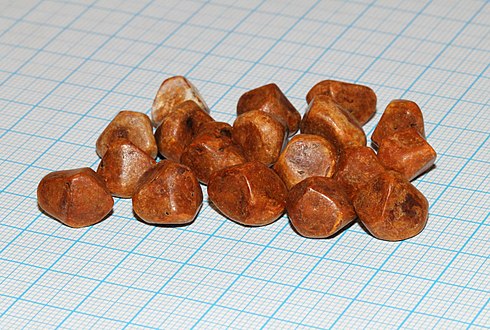
Cholesterol stones vary from light yellow to dark green or brown or chalk white and are oval, usually solitary, between 2 and 3 cm long, each often having a tiny, dark, central spot. To be classified as such, they must be at least 80% cholesterol by weight (or 70%, according to the Japanese classification system).[37] Between 35% and 90% of stones are cholesterol stones.[3]
Pigment stones
Bilirubin ("pigment", "black pigment") stones are small, dark (often appearing black), and usually numerous. They are composed primarily of bilirubin (insoluble bilirubin pigment polymer) and calcium (calcium phosphate) salts that are found in bile. They contain less than 20% of cholesterol (or 30%, according to the Japanese classification system).[37] Between 2% and 30% of stones are bilirubin stones.[3]
Mixed stones
Mixed (
Gallstones can vary in size and shape from as small as a grain of sand to as large as a golf ball.[38] The gallbladder may contain a single large stone or many smaller ones. Pseudoliths, sometimes referred to as sludge, are thick secretions that may be present within the gallbladder, either alone or in conjunction with fully formed gallstones.
-
 Gallbladder opened to show small cholesterol gallstones
 -
 X-ray microtomograph of a gallstone
 -
 The large, yellow stone is largely cholesterol, while the green-to-brown stones are mostly composed of bile pigments
 -
CT images of gallstones
-
 Large gallstone
 -
 Numerous small gallstones made up largely of cholesterol


Diagnosis
Diagnosis is typically confirmed by
On abdominal ultrasound, sinking gallstones usually have posterior acoustic shadowing. In floating gallstones, reverberation echoes (or comet-tail artifact) is seen instead in a clinical condition called adenomyomatosis. Another sign is wall-echo-shadow (WES) triad (or double-arc shadow) which is also characteristic of gallstones.[39]
A positive Murphy's sign is a common finding on physical examination during a gallbladder attack.
-
 A 1.9 cm gallstone impacted in the neck of the gallbladder and leading to cholecystitis as seen onultrasound. There is 4 mm gall bladder wall thickening.
 -
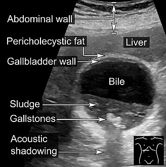 Biliary sludge and gallstones. There is borderline thickening of the gallbladder wall.
 -
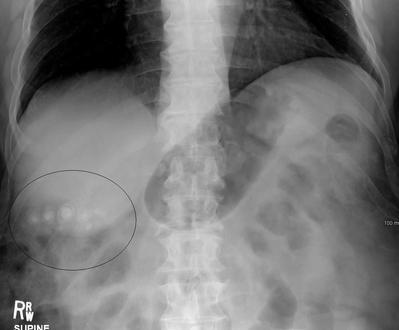 Gallstones as seen on plain X-ray
 -
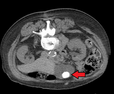 Large gallstone as seen on CT
 -
A normal gallbladder on ultrasound with bowel peristalsis creating the false appearance of stones


Prevention
Maintaining a healthy weight by getting sufficient exercise and eating a healthy diet that is high in fiber may help prevent gallstone formation.[2]
Ursodeoxycholic acid (UDCA) appears to prevent formation of gallstones during weight loss. A high fat diet during weight loss also appears to prevent gallstones.[40]
Treatment
Lithotripsy
Extracorporeal shock wave lithotripsy is a non-invasive method to manage gallstones that uses high-energy sound waves to disintegrate them first applied in January 1985.[41][42] Side effects of extracorporeal shock wave lithotripsy include biliary pancreatitis and liver haematoma.[43] The term is derived from the Greek words meaning 'breaking (or pulverizing) stones': litho- + τρίψω, tripso).
Surgical
Cholecystectomy (gallbladder removal) has a 99% chance of eliminating the recurrence of cholelithiasis. The lack of a gallbladder has no negative consequences in most people, however 10 to 15% of people develop postcholecystectomy syndrome,[44] which may cause nausea, indigestion, diarrhea, and episodes of abdominal pain.[45]
There are two surgical options for cholecystectomy:
- Open cholecystectomy is performed via an abdominal incision (laparotomy) below the lower right ribs. Recovery typically requires 3–5 days of hospitalization, with a return to normal diet a week after release and to normal activity several weeks after release.[12]
- Laparoscopic cholecystectomy, introduced in the 1980s, is performed via three to four small puncture holes for a camera and instruments. Post-operative care typically includes a same-day release or a one-night hospital stay, followed by a few days of home rest and pain medication.[12] Perforation of the gall bladder is not uncommon—it has been reported in the range of 10% to 40%. Unretrieved gallstone spillage has been reported as 6% to 30%, but gallstones that are not retrieved rarely cause complications (0.08%–0.3%).[46]
Obstruction of the common bile duct with gallstones can sometimes be relieved by endoscopic retrograde sphincterotomy (ERS) following endoscopic retrograde cholangiopancreatography (ERCP).[47]
Medical
The medications ursodeoxycholic acid (UDCA) and chenodeoxycholic acid (CDCA) have been used in treatment to dissolve gallstones.[48][49] A 2013 meta-analysis concluded that UDCA or higher dietary fat content appeared to prevent formation of gallstones during weight loss.[40] Medical therapy with oral bile acids has been used to treat small cholesterol stones, and for larger cholesterol gallstones when surgery is either not possible or unwanted. CDCA treatment can cause diarrhea, mild reversible hepatic injury, and a small increase in the plasma cholesterol level.[49] UDCA may need to be taken for years.[47]
Use in traditional medicine
Gallstones can be a valued by-product of animals butchered for meat because of their use as an antipyretic and antidote in the traditional medicine of some cultures, particularly traditional Chinese medicine. The most highly prized gallstones tend to be sourced from old dairy cows, termed calculus bovis or niu-huang (yellow thing of cattle) in Chinese. Some slaughterhouses carefully scrutinize workers for gallstone theft.[50]
See also
References
- ^ ISBN 978-0-7020-5483-9.
- ^ a b c d e f g h i j k l m n o p q r s t u v w x y z aa "Gallstones". NIDDK. November 2013. Archived from the original on 28 July 2016. Retrieved 27 July 2016.
- ^ PMID 26455113.
- ^ PMID 27307785.
- ^ PMID 25473723.
- ^ a b "Complications". nhs.uk. Retrieved 13 May 2018.
- ^ "Treatment for Gallstones". National Institute of Diabetes and Digestive and Kidney Diseases. November 2017.
- PMID 26063472.
- PMID 25530442.)
{{cite journal}}:|author1=has generic name (help)CS1 maint: numeric names: authors list (link - ISBN 978-1-4419-6999-6. Archivedfrom the original on 2016-08-15.
- S2CID 26695806.
- ^ a b c National Institute of Diabetes and Digestive and Kidney Diseases (2007). "Gallstones" (PDF). Bethesda, Maryland: National Digestive Diseases Information Clearinghouse, National Institutes of Health, United States Department of Health and Human Services. Archived from the original (PDF) on 2010-12-05. Retrieved 2010-11-06.
- ^ Heuman DM, Mihas AA, Allen J (2010). "Cholelithiasis". Omaha, Nebraska: Medscape (WebMD). Archived from the original on 2010-11-20. Retrieved 2010-11-06.
- S2CID 22457009.
- ^ a b "Gallstones (Cholelithiasis) Clinical Presentation: History, Physical Examination". emedicine.medscape.com. Archived from the original on 2016-11-14. Retrieved 2016-11-14.
- ^ "Symptoms & Causes of Gallstones | NIDDK". National Institute of Diabetes and Digestive and Kidney Diseases. Retrieved 2022-06-28.
- S2CID 43696188.
- ISBN 978-0-06-198079-4.
- ^ a b Afdhal N, Zakko S (Sep 2022). "Gallstones: Epidemiology, risk factors and prevention". UpToDate. Retrieved 2023-05-26.
- S2CID 22785223.
- S2CID 40558696.
- PMID 9013440. Archived from the originalon 2008-07-20. Retrieved 2010-11-06.
- ISBN 978-0-309-09169-5.
- PMID 15888792.
- S2CID 11355563.
- ^ NHS Choices. "Should you lose weight fast? – Live Well—NHS Choices". www.nhs.uk. Archived from the original on 2016-02-16. Retrieved 2016-02-16.
- ^ Office of the Commissioner. "Safety Information—Xenical (orlistat) capsules". www.fda.gov. Archived from the original on 2016-06-11. Retrieved 2016-06-18.
- PMID 28186337.
- PMID 7410545.
- ^ Endocrine and Metabolic Disorders: Cutaneous Porphyrias, pp. 63–220 in Beers, Porter and Jones (2006)
- ^ Thunell S (2008). "Endocrine and Metabolic Disorders: Cutaneous Porphyrias". Whitehouse Station, New Jersey: Merck Sharp & Dohme Corporation. Archived from the original on 2020-03-12. Retrieved 2010-11-07.
- S2CID 20833380.
- S2CID 25636425.
- ^ PMID 22910758.
- ^ Experimental investigation of the flow of bile in patient specific cystic duct models M Al-Atabi, SB Chin..., Journal of biomechanical engineering, 2010
- ^ Channa NA, Khand FD, Khand TU, Leghari MH, Memon AN (2007). "Analysis of human gallstones by Fourier Transform Infrared (FTIR)". Pakistan Journal of Medical Sciences. 23 (4): 546–50. Archived from the original on 2011-08-24. Retrieved 2010-11-06.
- ^ PMID 12950109.
- ^ Gallstones—Cholelithiasis; Gallbladder attack; Biliary colic; Gallstone attack; Bile calculus; Biliary calculus Archived 2011-02-07 at the Wayback Machine Last reviewed: July 6, 2009. Reviewed by: George F. Longstreth. Also reviewed by David Zieve
- PMID 3309915.
- ^ PMID 24321208.
- ^ "Gallstone Disease Treatment". Johns Hopkins Medicine. Retrieved 2021-09-25.
- PMID 15827443.
- PMID 15827443.
- ^ Jensen (2010). "Postcholecystectomy syndrome". Omaha, Nebraska: Medscape (WebMD). Archived from the original on 2010-12-23. Retrieved 2011-01-20.
- PMID 30969724.
- PMID 14970293.
- ^ a b National Health Service (2010). "Gallstones — Treatment". NHS Choices: Health A-Z—Conditions and treatments. London: National Health Service. Archived from the original on 2010-11-14. Retrieved 2010-11-06.
- PMID 4580472.
- ^ PMID 2672842.
- ^ "Interview with Darren Wise. Transcrip". Omaha, Nebraska: Medscape (WebMD). Archived from the original on 2010-11-21. Retrieved 2010-11-06.
External links
- "Gallstones". MedlinePlus. U.S. National Library of Medicine.