MAPK14
Ensembl | |||||||||
|---|---|---|---|---|---|---|---|---|---|
| UniProt | |||||||||
| RefSeq (mRNA) | |||||||||
| RefSeq (protein) | |||||||||
| Location (UCSC) | Chr 6: 36.03 – 36.11 Mb | Chr 17: 28.91 – 28.97 Mb | |||||||
| PubMed search | [3] | [4] | |||||||
| View/Edit Human | View/Edit Mouse |
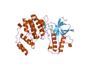
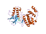
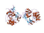
Mitogen-activated protein kinase 14, also called p38-α, is an enzyme that in humans is encoded by the MAPK14 gene.[5]
MAPK14 encodes p38α mitogen-activated protein kinase (MAPK) which is the prototypic member of the p38 MAPK family. p38 MAPKs are also known as stress-activated
Structure
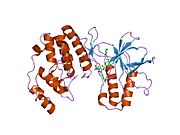
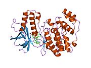
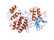
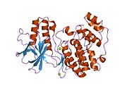
MAPK14 is a 41 kDa protein composed of 360 amino acids.[8][9]
Function
The protein encoded by this gene is a member of the
p38α MAPK is ubiquitously expressed in many cell types, in contrast, p38β MAPK is highly expressed in brain and lung, p38γ MAPK mostly in skeletal muscle and nerve system, and p38δ MAPK in uterus and pancreas.[11][12] Like all MAP kinases, p38α MAPK has 11 conserved domains (Domains I to XI) and a Thr-Gly-Tyr (TGY) dual phosphorylation motif. Activation of p38 MAPK pathway has been implicated in a variety of stress response in addition to inflammation, including osmotic shock, heat, and oxidative stress.[11][13][14] The canonical pathway for p38 MAPK activation involve a cascade of protein kinases, including MAP3K such as MEKK1, 2, 3 and 4, TGFβ-activated kinase (TAK1), TAO1-3, mixed-lineage kinase 2/3 (MLK2/3), and apoptosis signal-regulating kinase 1/2 (ASK1/2), as well as MAP2Ks, such as MKK3, 6 and 4. MAP2K mediated phosphorylation of the TGY motif results in conformational change of p38 MAPK which allows kinase activation and accessibility to substrates.[15] In addition, TAK1-binding protein 1 (TAB1) and ZAP70 can induce p38 MAPK via non-canonical autophosphorylation.[16][17][18] Furthermore, acetylation of p38 MAPK at lys-53 of the ATP-binding pocket also enhances p38 MAPK activity during cellular stress[19] Under basal conditions, p38α MAPK is detected in both the nucleus and the cytoplasm. One of the consequences of p38 MAPK activation is translocation into the nucleus.[20] involving both p38 MAPK phosphorylation and microtubule- and dynein-dependent process.[21] In addition, one substrate of p38 MAPK, MAP kinase-activated protein kinase 2 (MAPAK2 or MK2) can modulate and direct p38α MAPK localization to cytosole via direct interaction.[22] p38α MAPK activation can be reversed by dephosphorylation of the TGY motif carried out by protein phosphatases, including ser-thr protein phosphatases (PPs), protein tyrosine phosphatases (PTP), and dual-specificity phosphatases (DUSP). For example, ser/thr phosphatases PP2Cα/β suppress activity of p38s MAPK through direct interaction as well as suppression of MKKs/TAK1 in mammalian cells.[23][24] Hematopoietic PTP (HePTP) and striatal-enriched phosphatase (STEP) bind to MAPKs through a kinase-interaction motif (KIM) and inactivates them by dephosphorylating the phosphotyrosine residue in their activation loop.[25][26][27] DUSPs, which have a docking domain to MAPKs and dual-specific phosphatase activity, can also bind to p38 MAPKs and dephosphorylate of both phosphotyrosine and phosphothreonine residues.[15] In addition to these phosphatases, other molecular components such as Hsp90-Cdc37 chaperone complex can also modulate p38 MAPK autophosphorylation activity and prevents non-canonical activation.[28]
p38α MAPK is implicated in cell survival/apoptosis, proliferation, differentiation, migration, mRNA stability, and inflammatory response in different cell types through variety of different target molecules[29] MK2 is one of the well-studied downstream targets of p38α MAPK. Their downstream substrates include small heat shock protein 27 (HSP27), lymphocyte-specific protein1 (LSP1), cAMP response element-binding protein (CREB), cyclooxygenase 2 (COX2), activating transcription factor 1 (ATF1), serum response factor (SRF), and mRNA-binding protein tristetraprolin (TTP)[20][30] In addition to protein kinases, many transcription factors are downstream targets of p38α MAPK, including ATF1/2/6, c-Myc, c-FOS, GATA4, MEF2A/C, SRF, STAT1, and CHOP[31][32][33][34]
Role in cardiovascular system
p38α MAPK constitutes the main p38 MAPK activity in heart. During cardiomyocyte maturation in new born mouse heart, p38α MAPK activity can regulate myocyte cytokinesis and promote cell cycle exit.[35] while inhibition of p38 MAPK activity leads to induction of mitosis in both adult and fetal cardiomyocyte.[36][37] Therefore, p38 MAPK is associated with cell-cycle arrest in mammalian cardiomyocytes and its inhibition may represent a strategy to promote cardiac regeneration in response to injury. In addition, p38α MAPK induction promotes myocyte apoptosis.[38][39] via downstream targets STAT1, CHOP, FAK, SMAD, cytochrome c, NF-κB, PTEN, and p53.[40][41][42][43][44][45][46] p38 MAPK can also target IRS-1 mediated AKT signaling and promotes myocyte death under chronic insulin stimulation.[47] Inhibition of p38 MAPK activity confers cardioprotection against ischemia reperfusion injury in heart[48][49] However, some reports demonstrated that p38 MAPK also involves in anti-apoptotic effect via phosphorylation of αβ-Crystallin or induction of Pim-3 during early response to oxidative stress or anoxic preconditioning respectively[50][51][52] Both p38α MAPK and p38β MAPK appear to have an opposite role in apoptosis.[53] Whereas p38α MAPK has a pro-apoptotic role via p53 activation, p38β MAPK has a pro-survival role via inhibition of ROS formation.[54][55] In general, chronic activation of p38 MAPK activity is viewed as pathological and pro-apoptotic, and inhibition of p38 MAPK activity is in clinical evaluation as a potential therapy to mitigate acute injury in ischemic heart failure.[56] p38 MAPK activity is also implicated in cardiac hypertrophy which is a significant feature of pathological remodeling in the diseased hearts and a major risk factor for heart failure and advert outcome. Most in vitro evidence supports that p38 MAPK activation promotes cardiomyocyte hypertrophy.[53][57][58][59] However, in vivo evidence suggest that chronic activation of p38 MAPK activity triggers restrictive cardiomyopathy with limited hypertrophy,[60] while genetic inactivation p38α MAPK in mouse heart results in an elevated cardiac hypertrophy in response to pressure overload[61][62] or swimming exercise.[63] Therefore, the functional role of p38 MAPK in cardiac hypertrophy remains controversial and yet to be further elucidated.
Interactions
MAPK14 has been shown to
Notes
PMID 26390817 . |
References
- ^ a b c GRCh38: Ensembl release 89: ENSG00000112062 – Ensembl, May 2017
- ^ a b c GRCm38: Ensembl release 89: ENSMUSG00000053436 – Ensembl, May 2017
- ^ "Human PubMed Reference:". National Center for Biotechnology Information, U.S. National Library of Medicine.
- ^ "Mouse PubMed Reference:". National Center for Biotechnology Information, U.S. National Library of Medicine.
- S2CID 4306822.
- PMID 7914033.
- PMID 7693711.
- PMID 23965338.
- ^ "Mitogen-activated protein kinase 14". Cardiac Organellar Protein Atlas Knowledgebase (COPaKB).
- ^ "Entrez Gene: MAPK14 mitogen-activated protein kinase 14".
- ^ PMID 10676842.
- PMID 22046431.
- S2CID 33514114.
- PMID 11274345.
- ^ S2CID 9018714.
- S2CID 25004892.
- ^ S2CID 93896233.
- PMID 12829618.
- PMID 21444723.
- ^ PMID 15686620.
- S2CID 38862126.
- PMID 9768359.
- PMID 9824284.
- PMID 9707433.
- PMID 9857190.
- PMID 10206983.
- PMID 9624114.
- PMID 20299663.
- PMID 23896688.
- PMID 20959622.
- PMID 7535770.
- S2CID 678541.
- PMID 2997391.
- PMID 17699753.
- PMID 16889791.
- PMID 17032753.
- PMID 15870258.
- PMID 14749328.
- S2CID 21917525.
- PMID 16631627.
- PMID 11574421.
- PMID 19616567.
- PMID 22367504.
- S2CID 12670283.
- PMID 11309387.
- PMID 19237210.
- PMID 24159000.
- PMID 10190877.
- PMID 15808838.
- PMID 18420382.
- PMID 19505587.
- S2CID 10718645.
- ^ PMID 9442057.
- PMID 16407188.
- PMID 20724307.
- PMID 21062627.
- PMID 14654364.
- PMID 9584192.
- PMID 9314533.
- PMID 11593045.
- PMID 15572667.
- PMID 12639989.
- PMID 18195174.
- ^ PMID 11042204.
- PMID 7535770.
- ^ PMID 11279118.
- PMID 9207092.
- ^ S2CID 4410763.
- PMID 10747897.
- ^ PMID 11359773.
- ^ PMID 11157753.
- PMID 10391943.
- PMID 11489891.
- PMID 11278799.
- S2CID 41006480.
- PMID 10644717.
- PMID 11788583.
- ^ PMID 12697810.
- PMID 8622669.
- PMID 8626699.
- PMID 11551945.
- PMID 9858528.
- PMID 10330143.
- PMID 9792677.
- S2CID 4427026.
Further reading
- Ben-Levy R, Hooper S, Wilson R, Paterson HF, Marshall CJ (Sep 1998). "Nuclear export of the stress-activated protein kinase p38 mediated by its substrate MAPKAP kinase-2". Current Biology. 8 (19): 1049–57. PMID 9768359.
- Bradham C, McClay DR (Apr 2006). "p38 MAPK in development and cancer". Cell Cycle. 5 (8): 824–8. PMID 16627995.
- P. Sankaranarayanan, T. E. Schomay, K. A. Aiello, O. Alter (April 2015). "Tensor GSVD of Patient- and Platform-Matched Tumor and Normal DNA Copy-Number Profiles Uncovers Chromosome Arm-Wide Patterns of Tumor-Exclusive Platform-Consistent Alterations Encoding for Cell Transformation and Predicting Ovarian Cancer Survival". PLOS ONE. 10 (4): e0121396. .
External links
- MAP Kinase Resource Archived 2021-04-15 at the Wayback Machine.
- Overview of all the structural information available in the PDB for UniProt: Q16539 (Mitogen-activated protein kinase 14) at the PDBe-KB.


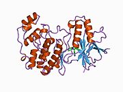

![1di9: The structure of p38 mitogen-activated protein kinase in complex with 4-[3-methylsulfanylanilino]-6,7-dimethoxyquinazoline](http://upload.wikimedia.org/wikipedia/commons/thumb/1/18/PDB_1di9_EBI.jpg/180px-PDB_1di9_EBI.jpg)


