Evolution of nervous systems

Source: Wikipedia, the free encyclopedia.

The evolution of nervous systems dates back to the first development of

epithelia, contractile muscles and coordinating & sensitive neurons for it in their outer layer.[2][3]

Simple

phylogenetic tree of life is still disputed.[5][6][7] Further cephalization and nerve cord (ventral and dorsal) evolution occurred many times independently in bilaterians.[5]

Neural precursors

NUCB2, that are precursors of the neuropeptides phoenixin and nesfatin-1 respectively have been found to have deep homology across all lineages that preceded creatures with central nervous systems, bilaterians, cnidarians, ctenophores, and sponges as well as in choanoflagellates.[11][12]

Sponges

Sponges have no cells connected to each other by synaptic junctions, that is, no neurons, and therefore no nervous system. They do, however, have homologs of many genes that play key roles in synaptic function. Recent studies have shown that sponge cells express a group of proteins that cluster together to form a structure resembling a postsynaptic density (the signal-receiving part of a synapse).[13] However, the function of this structure is currently unclear. Although sponge cells do not show synaptic transmission, they do communicate with each other via calcium waves and other impulses, which mediate some simple actions such as whole-body contraction.[14] Other ways sponge cells communicate with neighboring cells is through vesicular transport across highly dense regions of the cell membranes. These vesicles carry ions and other signaling molecules, but contain no true synaptic function.[15]

Nerve nets

comb jellies, and related animals have diffuse nerve nets rather than a central nervous system. In most jellyfish the nerve net is spread more or less evenly across the body; in comb jellies it is concentrated near the mouth. The nerve nets consist of sensory neurons that pick up chemical, tactile, and visual signals, motor neurons that can activate contractions of the body wall, and intermediate neurons that detect patterns of activity in the sensory neurons and send signals to groups of motor neurons as a result. In some cases groups of intermediate neurons are clustered into discrete ganglia.[16]

The development of the nervous system in

bilaterians, radiata only have two primordial cell layers, endoderm and ectoderm. Neurons are generated from a special set of ectodermal precursor cells, which also serve as precursors for every other ectodermal cell type.[17]

Nerve cords

A rod-shaped body contains a digestive system running from the mouth at one end to the anus at the other. Alongside the digestive system is a nerve cord with a brain at the end, near to the mouth.
Nervous system of a bilaterian animal, in the form of a nerve cord with a "brain" at the front

The vast majority of existing animals are bilaterians, meaning animals with left and right sides that are approximate mirror images of each other. All bilateria are thought to have descended from a common wormlike ancestor that appeared in the Cryogenian period, 700–650 million years ago.[18] The fundamental bilaterian body form is a tube with a hollow gut cavity running from mouth to anus, and a nerve cord with an especially large ganglion at the front, called the "brain".
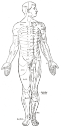
Area of the human body surface innervated by each spinal nerve

Even

mammals, including humans, show the segmented bilaterian body plan at the level of the nervous system. The spinal cord contains a series of segmental ganglia, each giving rise to motor and sensory nerves that innervate a portion of the body surface and underlying musculature. On the limbs, the layout of the innervation pattern is complex, but on the trunk it gives rise to a series of narrow bands. The top three segments belong to the brain, giving rise to the forebrain, midbrain, and hindbrain.[19]

Bilaterians can be divided, based on events that occur very early in embryonic development, into two groups (

molluscs, and numerous types of worms. There is a basic difference between the two groups in the placement of the nervous system within the body: protostomes possess a nerve cord on the ventral (usually bottom) side of the body, whereas in deuterostomes the nerve cord is on the dorsal (usually top) side. In fact, numerous aspects of the body are inverted between the two groups, including the expression patterns of several genes that show dorsal-to-ventral gradients. Some anatomists now consider that the bodies of protostomes and deuterostomes are "flipped over" with respect to each other, a hypothesis that was first proposed by Geoffroy Saint-Hilaire for insects in comparison to vertebrates. Thus insects, for example, have nerve cords that run along the ventral midline of the body, while all vertebrates have spinal cords that run along the dorsal midline.[21] But recent molecular data from different protostomes and deuterostomes reject this scenario and suggest that nerve cords independently evolved in both.[22]

Annelida

Photoreceptors on the animal's eyespots provide sensory information on light and dark.[23]

Nematoda

The nervous system of one very small worm, the

hermaphrodites, have different numbers of neurons and groups of neurons that perform sex-specific functions. In C. elegans, males have exactly 383 neurons, while hermaphrodites have exactly 302 neurons.[24]

Arthropods

Internal anatomy of a spider, showing the nervous system in blue

olfaction and pheromone
sensation. The sensory information from these organs is processed by the brain.

In insects, many neurons have cell bodies that are positioned at the edge of the brain and are electrically passive—the cell bodies serve only to provide metabolic support and do not participate in signalling. A protoplasmic fiber runs from the cell body and branches profusely, with some parts transmitting signals and other parts receiving signals. Thus, most parts of the insect brain have passive cell bodies arranged around the periphery, while the neural signal processing takes place in a tangle of protoplasmic fibers called neuropil, in the interior.[26]

Evolution of central nervous systems

Evolution of the human brain

There has been a gradual increase in

chimpanzee. It is proposed that they evolved from H. erectus as a case of insular dwarfism. In spite of their threefold smaller brain there is evidence that H. floresiensis used fire and made stone tools as sophisticated as those of their proposed ancestor, H. erectus.[28] Iain Davidson summarizes the opposite evolutionary constraints on human brain size as "As large as you need and as small as you can".[29] The human brain has evolved around the metabolic, environmental, and social needs that the species has dealt with throughout its existence. As hominid species evolved with increased brain size and processing power, the overall metabolic need increased. Compared to chimpanzees, humans consume more calories from animals than from plants. While not certain, studies have shown that this shift in diet is due to the increased need for the fatty acids more readily found in animal products[citation needed]. These fatty acids are essential for brain maintenance and development. Other factors to consider are the need for social interaction and how hominids have interacted with their environments over time.[30]

Brain evolution can be studied using

paleoneurology
.

See also

References

  1. ^ "nervous system | Definition, Function, Structure, & Facts". Encyclopædia Britannica. Retrieved 2021-04-07.
  2. ^ Arendt, D. (2021). Elementary Nervous Systems. Philosophical Transactions of the Royal Society B: Biological Sciences, 376 (1821), 20200347. https://doi.org/10.1098/rstb.2020.0347
  3. ^ Arendt, D., Benito-Gutierrez, E., Brunet, T., & Marlow, H. (2015). Gastric pouches and the mucociliary sole: Setting the stage for nervous system evolution. Philosophical Transactions of the Royal Society B: Biological Sciences, 370 (1684), 20150286. https://doi.org/10.1098/rstb.2015.0286
  4. ^ Jékely, G., Paps, J. & Nielsen, C. The phylogenetic position of ctenophores and the origin(s) of nervous systems. EvoDevo 6, 1 (2015). https://doi.org/10.1186/2041-9139-6-1
  5. ^ a b Moroz, L. L., Romanova, D. Y., & Kohn, A. B. (2021). Neural versus alternative integrative systems: Molecular insights into origins of neurotransmitters. Philosophical Transactions of the Royal Society B: Biological Sciences, 376 (1821), 20190762. https://doi.org/10.1098/rstb.2019.0762
  6. ^ Musser, J. M., Schippers, K. J., Nickel, M. et al. (2021). Profiling cellular diversity in sponges informs animal cell type and nervous system evolution. Science, 374 (6568), 717–723. https://doi.org/10.1126/science.abj2949
  7. ^ Hayakawa, E., Guzman, C., Horiguchi, O. et al. Mass spectrometry of short peptides reveals common features of metazoan peptidergic neurons. Nat Ecol Evol (2022). https://doi.org/10.1038/s41559-022-01835-7
  8. .
  9. .
  10. .
  11. .
  12. ^ Callier V (2022-06-03). "Brain-Signal Proteins Evolved Before Animals Did". Quanta Magazine. Retrieved 2022-06-03.
  13. PMID 17551586
    .
  14. .
  15. .
  16. .
  17. .
  18. .
  19. .
  20. .
  21. .
  22. .
  23. .
  24. ^ "Specification of the nervous system". Wormbook.
  25. .
  26. ^ Chapman, p. 546
  27. ^ Ko KH (2016). "Origins of human intelligence: The chain of tool-making and brain evolution" (PDF). Anthropological Notebooks. 22 (1): 5–22.
  28. S2CID 26441
    .
  29. .
  30. .