
Cutaneous squamous-cell carcinoma
| Cutaneous squamous-cell carcinoma | |
|---|---|
| Other names | Squamous-cell carcinoma of the skin, squamous-cell skin cancer, epidermoid carcinoma, squamous-cell epithelioma of the skin |
| Treatment | Surgical removal, radiotherapy, chemotherapy, immunotherapy[2][7] |
| Prognosis | Usually good[5] |
| Frequency | 2.2 million (2015)[8] |
| Deaths | 51,900 (2015)[9] |
Cutaneous squamous-cell carcinoma (cSCC), also known as squamous-cell carcinoma of the skin or squamous-cell skin cancer, is one of the three principal types of skin cancer, alongside basal-cell carcinoma and melanoma.[10] cSCC typically presents as a hard lump with a scaly surface, though it may also present as an ulcer.[1] Onset and development often occurs over several months.[4] Compared to basal cell carcinoma, cSCC is more likely to spread to distant areas.[11] When confined to the epidermis, the outermost layer of the skin, the pre-invasive or in situ form of cSCC is termed Bowen's disease.[12][13]
The most significant risk factor for cSCC is extensive lifetime exposure to
Research, both in vivo and in vitro, indicates a crucial role for the
Preventive measures against cSCC include minimizing exposure to ultraviolet radiation and the use of
As of 2015, approximately 2.2 million individuals globally were living with cSCC at any given time,[8] constituting about 20% of all skin cancer cases.[21] In the United States, approximately 12% of males and 7% of females are diagnosed with cSCC at some point in their lives.[2] While prognosis remains favorable in the absence of metastasis, upon distant spread the five-year survival rate is markedly reduced to ~34%.[4][5] In 2015, global deaths attributed to cSCC numbered around 52,000.[9] The average age at diagnosis is approximately 66 years.[4] Following successful treatment of an initial cSCC lesion, there is a substantial risk of developing subsequent lesions.[2] As of 2015, about 2.2 million people worldwide have cSCC at any given time. About 20% of all skin cancer cases consist of cSCC. About 12% of males and 7% of females in the United States develop cSCC at some point in time. While prognosis is usually good, when distant spread occurs
Signs and symptoms

SCC of the skin begins as a small nodule and as it enlarges the center becomes necrotic and sloughs and the nodule turns into an ulcer, and generally are developed from an actinic keratosis. Once keratinocytes begin to grow uncontrollably, they have the potential to become cancerous and produce cutaneous squamous-cell carcinoma.[22]
- The lesion caused by cSCC is often asymptomatic
- Ulcer or reddish skin plaque that is slow growing
- Intermittent bleeding from the tumor, especially on the lip
- The clinical appearance is highly variable
- Usually the tumor presents as an ulcerated lesion with hard, raised edges
- The tumor may be in the form of a hard plaque or a papule, often with an opalescent quality, with tiny blood vessels
- The tumor can lie below the level of the surrounding skin, and eventually ulcerates and invades the underlying tissue
- The tumor commonly presents on sun-exposed areas (e.g. back of the hand, scalp, lip, and superior surface of pinna)
- On the lip, the tumor forms a small ulcer, which fails to heal and bleeds intermittently
- Evidence of chronic skin photodamage, as in multiple actinic keratoses (solar keratoses)
- The tumor grows relatively slowly
Spread
- Unlike basal-cell carcinoma (BCC), squamous-cell carcinoma (SCC) has a higher risk of metastasis.
- Risk of metastasis is higher clinically in SCC arising in scars, on the lower lips, ears, or mucosa, and occurring in immunosuppressed and solid organ transplant patients. Risk of metastasis is also higher in SCC that are > 2 cm in diameter, growth into the fat layer and along nerves, presence of lymphovascular invasion, poorly differentiated cell architecture on histology, or thickness greater than 6 mm.[23][24][25]
Causes
Cutaneous squamous-cell carcinoma is the second-most common
cSCC represents about 20% of the non-melanoma skin cancers; 80-90% of cSCCs with metastatic potential are located on the head and neck.[29]
Tobacco smoking also increases the risk for cutaneous squamous-cell carcinoma.[14][30]
The vast majority of cSCC cases are located on exposed skin, and are often the result of ultraviolet exposure. cSCC usually occurs on portions of the body commonly exposed to the sun; the face, ears, neck, hands, or arms. The primary sign is a growing bump that may have a rough, scaly surface, and flat, reddish patches. Unlike basal-cell carcinoma, cSCC carries a higher risk of metastasis than does basal-cell carcinoma, and may spread to the regional lymph nodes,[31]
Erythroplasia of Queyrat (SCC in situ of the
Genetically, cSCC tumors harbor high frequencies of NOTCH and p53 mutations as well as less frequent alterations in histone acetyltransferase EP300, subunit of the SWI/SNF chromatin remodeling complex PBRM1, DNA-repair deubiquitinase USP28, and NF-κB signaling regulator CHUK.[40]
Immunosuppression
People who have received solid organ transplants are at a significantly increased risk of developing squamous-cell carcinoma due to the use of chronic immunosuppressive medication.[41] While the risk of developing all skin cancers increases with these medications, this effect is particularly severe for cSCC, with hazard ratios as high as 250 being reported, versus 40 for basal cell carcinoma.[42] The incidence of cSCC development increases with time posttransplant.[43] Heart and lung transplant recipients are at the highest risk of developing cSCC due to more intensive immunosuppressive medications used.[44]
Cutaneous squamous-cell carcinoma in individuals on immunotherapy or who have lymphoproliferative disorders (e.g. leukemia) tend to be much more aggressive, regardless of their location.[45] The risk of cSCC, and non-melanoma skin cancers generally, varies with the immunosuppressive drug regimen chosen. The risk is greatest with calcineurin inhibitors like cyclosporine and tacrolimus, and least with mTOR inhibitors, such as sirolimus and everolimus. The antimetabolites azathioprine and mycophenolic acid have an intermediate risk profile.[46]
Diagnosis
Diagnosis is confirmed via skin biopsy of the tissue or tissues suspected to be affected by SCC. The pathological appearance of a squamous-cell cancer varies with the depth of the biopsy. For that reason, a biopsy including the subcutaneous tissue and basilar epithelium, to the surface is necessary for correct diagnosis. The performance of a shave biopsy (see skin biopsy) might not acquire enough information for a diagnosis. An inadequate biopsy might be read as actinic keratosis with follicular involvement. A deeper biopsy down to the dermis or subcutaneous tissue might reveal the true cancer. An excision biopsy is ideal, but not practical in most cases. An incisional or punch biopsy is preferred. A shave biopsy is least ideal, especially if only the superficial portion is acquired.[citation needed]
Histological characteristics
Histopathologically, the epidermis in cSCC in situ (Bowen's disease) will show hyperkeratosis and parakeratosis. There will also be marked acanthosis with elongation and thickening of the rete ridges. These changes will overly keratinocytic cells which are often highly atypical and may in fact have a more unusual appearance than invasive cSCC. The atypia spans the full thickness of the epidermis, with the keratinocytes demonstrating intense mitotic activity, pleomorphism, and greatly enlarged nuclei. They will also show a loss of maturity and polarity, giving the epidermis a disordered or "windblown" appearance.[citation needed]
Two types of multinucleated cells may be seen: the first will present as a multinucleated giant cell, and the second will appear as a dyskeratotic cell engulfed in the cytoplasm of a keratinocyte. Occasionally, cells of the upper epidermis will undergo vacuolization, demonstrating an abundant and strongly eosinophilic cytoplasm. There may be a mild to moderate lymphohistiocytic infiltrate detected in the upper dermis.[12]
-
 Histopathology of squamous-cell carcinoma in situ (black arrow), compared to normal skin, showing marked atypia.
 -
![Squamous-cell carcinoma in situ, showing prominent dyskeratosis and aberrant mitoses at all levels of the epidermis, along with marked parakeratosis.[12]](//upload.wikimedia.org/wikipedia/commons/thumb/b/ba/Micrograph_of_squamous_cell_carcinoma_in_situ_-_100x.jpg/367px-Micrograph_of_squamous_cell_carcinoma_in_situ_-_100x.jpg) Squamous-cell carcinoma in situ, showing prominent dyskeratosis and aberrant mitoses at all levels of the epidermis, along with marked parakeratosis.[12]
Squamous-cell carcinoma in situ, showing prominent dyskeratosis and aberrant mitoses at all levels of the epidermis, along with marked parakeratosis.[12]

![Squamous-cell carcinoma in situ, showing prominent dyskeratosis and aberrant mitoses at all levels of the epidermis, along with marked parakeratosis.[12]](/File:Micrograph_of_squamous_cell_carcinoma_in_situ_-_100x.jpg)
In situ disease
Bowen's disease is essentially equivalent to and used interchangeably with cSCC in situ, when not having invaded through the
-
![cSCC in situ, high magnification, demonstrating an intact basement membrane.[12]](//upload.wikimedia.org/wikipedia/commons/thumb/0/01/Micrograph_of_squamous_cell_carcinoma_in_situ_-_400x.jpg/299px-Micrograph_of_squamous_cell_carcinoma_in_situ_-_400x.jpg) cSCC in situ, high magnification, demonstrating an intact basement membrane.[12]
cSCC in situ, high magnification, demonstrating an intact basement membrane.[12] -
 cSCC in situ
 -
 cSCC in situ
 -
 cSCC in situ
 -
 cSCC in situ
![cSCC in situ, high magnification, demonstrating an intact basement membrane.[12]](/File:Micrograph_of_squamous_cell_carcinoma_in_situ_-_400x.jpg)
.jpg)
.jpg)
.jpg)
.jpg)
Erythroplasia of Queyrat is a particular type of Bowen's disease that can arise on the glans or prepuce in males,[32][33]: 733 [34]: 656 [35] and the vulva in females.[36] It mainly occurs in uncircumcised males,[36][49] over the age of 40.[39]
Invasive disease
In invasive cSCC, tumor cells infiltrate through the basement membrane. The infiltrate can be somewhat difficult to detect in the early stages of invasion: however, additional indicators such as full thickness epidermal atypia and the involvement of hair follicles can be used to facilitate the diagnosis. Later stages of invasion are characterized by the formation of nests of atypical tumor cells in the dermis, often with a corresponding inflammatory infiltrate.[12]
-
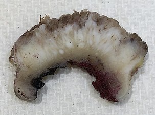 Gross slice of squamous-cell carcinoma of the skin
 -
![Superficially invasive cutaneous squamous-cell carcinoma. These lesions often do not show the marked pleomorphism and atypical nuclei of cSCC in situ, but manifest early keratinocyte invasion of the dermis.[12]](//upload.wikimedia.org/wikipedia/commons/thumb/f/f8/Micrograph_of_invasive_squamous_cell_carcinoma_-_150x.jpg/305px-Micrograph_of_invasive_squamous_cell_carcinoma_-_150x.jpg) Superficially invasive cutaneous squamous-cell carcinoma. These lesions often do not show the marked pleomorphism and atypical nuclei of cSCC in situ, but manifest early keratinocyte invasion of the dermis.[12]
Superficially invasive cutaneous squamous-cell carcinoma. These lesions often do not show the marked pleomorphism and atypical nuclei of cSCC in situ, but manifest early keratinocyte invasion of the dermis.[12] -
![High magnification demonstrates the pleomorphism of the invading keratinocytes[12]](//upload.wikimedia.org/wikipedia/commons/thumb/2/26/Micrograph_of_invasive_squamous_cell_carcinoma_-_200x.jpg/340px-Micrograph_of_invasive_squamous_cell_carcinoma_-_200x.jpg) High magnification demonstrates the pleomorphism of the invading keratinocytes[12]
High magnification demonstrates the pleomorphism of the invading keratinocytes[12] -
 Invasive nests with characteristic large celled centers. Ulceration (at left) is common in invasive cSCC.

![Superficially invasive cutaneous squamous-cell carcinoma. These lesions often do not show the marked pleomorphism and atypical nuclei of cSCC in situ, but manifest early keratinocyte invasion of the dermis.[12]](/File:Micrograph_of_invasive_squamous_cell_carcinoma_-_150x.jpg)
![High magnification demonstrates the pleomorphism of the invading keratinocytes[12]](/File:Micrograph_of_invasive_squamous_cell_carcinoma_-_200x.jpg)

Degree of differentiation
-
![Well-differentiated (yet invasive) cSCC, showing prominent keratinization. It may form pearl-like structures where dermal nests of keratinocytes attempt to mature in a layered fashion. Well-differentiated cSCC has slightly enlarged hyperchromatic nuclei with abundant amounts of cytoplasm. Intercellular bridges will frequently be visible.[12]](//upload.wikimedia.org/wikipedia/commons/thumb/3/3c/Micrograph_of_well-differentiated_and_invasive_squamous-cell_carcinoma.jpg/306px-Micrograph_of_well-differentiated_and_invasive_squamous-cell_carcinoma.jpg) Well-differentiated (yet invasive) cSCC, showing prominent keratinization. It may form pearl-like structures where dermal nests of keratinocytes attempt to mature in a layered fashion. Well-differentiated cSCC has slightly enlarged hyperchromatic nuclei with abundant amounts of cytoplasm. Intercellular bridges will frequently be visible.[12]
Well-differentiated (yet invasive) cSCC, showing prominent keratinization. It may form pearl-like structures where dermal nests of keratinocytes attempt to mature in a layered fashion. Well-differentiated cSCC has slightly enlarged hyperchromatic nuclei with abundant amounts of cytoplasm. Intercellular bridges will frequently be visible.[12] -
![Moderately differentiated lesions of invasive cSCC show much less organization and maturation with significantly less keratin formation.[12]](//upload.wikimedia.org/wikipedia/commons/thumb/f/f2/Micrograph_of_moderately_differentiated_and_invasive_squamous-cell_carcinoma.jpg/310px-Micrograph_of_moderately_differentiated_and_invasive_squamous-cell_carcinoma.jpg) Moderately differentiated lesions of invasive cSCC show much less organization and maturation with significantly less keratin formation.[12]
Moderately differentiated lesions of invasive cSCC show much less organization and maturation with significantly less keratin formation.[12] -
![Poorly differentiated, where attempts at keratinization are often no longer evident. This is a clear-cell squamous-cell carcinoma. The dysplastic cells infiltrated cords through the dermis. Poorly differentiated cSCC has greatly enlarged pleomorphic nuclei showing a high degree of atypia and frequent mitoses.[12]](//upload.wikimedia.org/wikipedia/commons/thumb/6/61/Micrograph_of_clear-cell_squamous-cell_carcinoma.jpg/296px-Micrograph_of_clear-cell_squamous-cell_carcinoma.jpg) Poorly differentiated, where attempts at keratinization are often no longer evident. This is a clear-cell squamous-cell carcinoma. The dysplastic cells infiltrated cords through the dermis. Poorly differentiated cSCC has greatly enlarged pleomorphic nuclei showing a high degree of atypia and frequent mitoses.[12]
Poorly differentiated, where attempts at keratinization are often no longer evident. This is a clear-cell squamous-cell carcinoma. The dysplastic cells infiltrated cords through the dermis. Poorly differentiated cSCC has greatly enlarged pleomorphic nuclei showing a high degree of atypia and frequent mitoses.[12] -
 Poorly differentiated clear-cell squamous-cell carcinoma. For this type of cSCC, immunostains will likely be required to classify it unless other areas of the tumor show obvious squamous-cell features such as seen here (arrow).
![Well-differentiated (yet invasive) cSCC, showing prominent keratinization. It may form pearl-like structures where dermal nests of keratinocytes attempt to mature in a layered fashion. Well-differentiated cSCC has slightly enlarged hyperchromatic nuclei with abundant amounts of cytoplasm. Intercellular bridges will frequently be visible.[12]](/File:Micrograph_of_well-differentiated_and_invasive_squamous-cell_carcinoma.jpg)
![Moderately differentiated lesions of invasive cSCC show much less organization and maturation with significantly less keratin formation.[12]](/File:Micrograph_of_moderately_differentiated_and_invasive_squamous-cell_carcinoma.jpg)
![Poorly differentiated, where attempts at keratinization are often no longer evident. This is a clear-cell squamous-cell carcinoma. The dysplastic cells infiltrated cords through the dermis. Poorly differentiated cSCC has greatly enlarged pleomorphic nuclei showing a high degree of atypia and frequent mitoses.[12]](/File:Micrograph_of_clear-cell_squamous-cell_carcinoma.jpg)

Prevention
Appropriate sun-protective clothing, use of broad-spectrum (UVA/UVB) sunscreen with at least SPF 50, and avoidance of intense sun exposure may prevent skin cancer.[50] A 2016 review of sunscreen for preventing cutaneous squamous-cell carcinoma found insufficient evidence to demonstrate whether it was effective.[51]
Management
Most cutaneous squamous-cell carcinomas are removed with surgery. A few selected cases are treated with
After removal of the cancer, closure of the skin for patients with a decreased amount of skin laxity involves a split-thickness skin graft. A donor site is chosen and enough skin is removed so that the donor site can heal on its own. Only the epidermis and a partial amount of dermis is taken from the donor site which allows the donor site to heal. Skin can be harvested using either a mechanical dermatome or Humby knife.[56]
Electrodessication and curettage (EDC) can be done on selected squamous-cell carcinoma of the skin. In areas where cSCC is known to be non-aggressive, and where the patient is not immunosuppressed, EDC[clarification needed] can be performed with good to adequate cure rate.[57]
Treatment options for cSCC in situ (Bowen's disease) include
High-risk squamous-cell carcinoma, as defined by that occurring around the eye, ear, or nose, is of large size, is poorly differentiated, and grows rapidly, requires more aggressive, multidisciplinary management.
Nodal spread:
- Surgical block dissection if palpable nodes or in cases of Marjolin's ulcers but the benefit of prophylactic block lymph node dissection with Marjolin's ulcers is not proven.
- Radiotherapy
- Adjuvant therapy may be considered in those with high-risk cSCC even in the absence of evidence for local metastasis. Imiquimod (Aldara) has been used with success for squamous-cell carcinoma in situ of the skin and the penis, but the morbidity and discomfort of the treatment is severe. An advantage is the cosmetic result: after treatment, the skin resembles normal skin without the usual scarring and morbidity associated with standard excision. Imiquimod is not FDA-approved for any squamous-cell carcinoma.
In general, squamous-cell carcinomas have a high risk of local recurrence, and up to 50% do recur.[58] Frequent skin exams with a dermatologist is recommended after treatment.
Prognosis
The long-term outcome of squamous-cell carcinoma is dependent upon several factors: the sub-type of the carcinoma, available treatments, location and severity, and various patient health-related variables (accompanying diseases, age, etc.). Generally, the long-term outcome is positive, with a metastasis rate of 1.9-5.2% and a mortality rate of 1.5-3.4%.[25][59][60]
When it does metastasize, the most commonly involved organs are the lungs, brain, bone and other skin locations.[61] Squamous-cell carcinoma occurring in immunosuppressed people (such as those with organ transplant, human immunodeficiency virus infection, or chronic lymphocytic leukemia) the risk of developing cSCC and having metastasis is much higher than the general population.[62]
One study found squamous-cell carcinoma of the penis had a much greater rate of mortality than some other forms of squamous-cell carcinoma, that is, about 23%,[63] although this relatively high mortality rate may be associated with possibly latent diagnosis of the disease due to patients avoiding genital exams until the symptoms are debilitating, or refusal to submit to a possibly scarring operation upon the genitalia.
Epidemiology

The incidence of cutaneous squamous-cell carcinoma continues to rise around the world. This is theorized to be due to several factors; including an aging population, a greater incidence of those who are immunocompromised and the increasing use of tanning beds.[25]
A recent study estimated that there are between 180,000 and 400,000 cases of cSCC in the United States in 2013.[65] Risk factors for cSCC varies with age, gender, race, geography, and genetics. The incidence of cSCC increases with age and with those 75 years or older eing at a 5-10 times increased risk of developing cSCC as compared with those who are younger than 55 years old.[25] Males are affected with cSCC at a ratio of 3:1 in comparison to females.[25] Those who have light skin, red or blonde hair and light colored eyes are also at increased risk.[25]
Squamous-cell carcinoma of the skin can be found on all areas of the body but is most common on frequently sun-exposed areas, such as the face, legs and arms.[66] Solid organ transplant recipients (heart, lung, liver, pancreas, among others) are also at a heightened risk of developing aggressive, high-risk cSCC. There are also a few rare congenital diseases predisposed to cutaneous malignancy. In certain geographic locations, exposure to arsenic in well water[67] or from industrial sources may significantly increase the risk of cSCC.[26]
Additional images
-
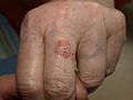
 Biopsy-proven cutaneous squamous-cell carcinoma
 -
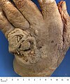 Squamous-cell carcinoma of the dorsum of the hand
 -
 cSCC in situ (Bowen's disease)
 -
 cSCC of the right upper cheek; lesion outlined in blue with a dashed line prior to biopsy
 -
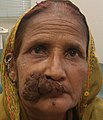
 Giant squamous cell carcinoma of the cheek


See also
References
- ^ ISBN 9780803626478. Archivedfrom the original on 2016-05-20.
- ^ PMID 26476255.
- ^ a b "Skin Cancer Treatment". National Cancer Institute. 21 June 2017. Archived from the original on 4 July 2017. Retrieved 2 July 2017.
- ^ ISBN 9780323448383. Archivedfrom the original on 29 August 2017. Retrieved 2 July 2017.
- ^ ISBN 978-9283204299.
- ^ PMID 22660875.
- ^ a b c "Skin Cancer Treatment". National Cancer Institute. 21 June 2017. Archived from the original on 4 July 2017. Retrieved 2 July 2017.
- ^ PMID 27733282.
- ^ PMID 27733281.
- ^ "Skin Cancer Treatment (PDQ®)". NCI. 2013-10-25. Archived from the original on 5 July 2014. Retrieved 30 June 2014.
- PMID 23084294.
- ^ license.
- ^ PMID 23794286.
- ^ a b "Basal and Squamous Cell Skin Cancer Risk Factors". American Cancer Society.
- JSTOR j.ctt1g69xq0.9.
- PMID 21199599.
- PMID 22044607.
- S2CID 221861310.
- ^ "NCI Dictionary of Cancer Terms". National Cancer Institute. 2011-02-02. Archived from the original on 9 November 2016. Retrieved 9 November 2016.
- ^ PMID 31254372.
- PMID 26219687.
- ^ H, Carlos (2022-09-10). "How serious is a squamous-cell carcinoma?". CuradermBCC. Archived from the original on 2022-09-10. Retrieved 2022-09-10.
- PMID 26762219.
- S2CID 245153567.
- ^ S2CID 259154802.
- ^ a b "MD Consult - Important Notice". Archived from the original on 3 March 2016. Retrieved 15 March 2017.
- ^ "Squamous Cell Carcinoma". American Academy of Dermatology. 25 December 2010. Archived from the original on 25 December 2010. Retrieved 15 March 2017.
- PMID 20935675.
- S2CID 245812900.
- PMID 11134217.
- ^ Wells JW, Goyal N, Najjar T, Monroe MM (28 June 2016). Meyers AD (ed.). "Cutaneous Squamous Cell Carcinoma: Practice Essentials, Background, Pathophysiology". Archived from the original on 6 April 2017. Retrieved 15 March 2017.
- ^ ISBN 978-1-4160-3185-7.
- ^ ISBN 978-0-07-138076-8.
- ^ ISBN 0-7216-2921-0.
- ^ ISBN 978-1-4160-2999-1.
- ^ ISBN 9781449619848.
- PMID 10951274.
- ^ Shah IA, Sangi SA, Abbasi SA (1998). "Bowen's Disease". Pakistan Journal of Ophthalmology. 14 (1): 37–38.
- ^ ISBN 978-3-540-00878-1.
- PMID 34272401.
- PMID 34272401.
- S2CID 40490951.
- PMID 21763561.
- PMID 26239556.
- ^ "Squamous Cell Carcinoma: What it Looks Like". SkinCancerNet. American Academy of Dermatology. 2008. Archived from the original on 2008-12-21.
- S2CID 25776283.
- ^ Khalyl-Mawad J (3 April 2021). "Pathology of Cutaneous Squamous Cell Carcinoma and Bowen Disease". Medscape. Updated: Jun 11, 2019
- ^ "Bowen's disease". National Health Service. 17 October 2017. Page last reviewed: 21 May 2019
- ISBN 0911910875.
- PMID 21199599.
- PMID 27455163.
- PMID 20393962.
- ^ Gross KG, Steinman HK, Rapini RP, eds. (1999). Mohs Surgery, Fundamentals and Techniques. Mosby.
- S2CID 231899301.
- S2CID 226231693.
- ^ [1], Hallock G. Squamous Cell Carcinoma Excision from Right Forearm with Split-Thickness Skin Graft from the Thigh. J Med Ins. 2020;2020(290.16) doi:https://jomi.com/article/290.16
- PMID 32931212.
- PMID 2921745.
- PMID 22545495.
- PMID 18617440.
- PMID 28344135.
- PMID 23818517.
- PMID 22386252.
- ^ "WHO Disease and injury country estimates". World Health Organization. 2009. Archived from the original on 2009-11-11. Retrieved Nov 11, 2009.
- PMID 23375456.
- ^ "Squamous Cell Carcinoma". T he Skin Cancer Foundation. 2018. Retrieved Dec 12, 2018.
- PMID 26231242.