
User:Flyer22 Frozen/Human brain
| Human brain | |
|---|---|
terminal vein, choroid vein, cerebellar veins | |
| Identifiers | |
| Latin | Cerebrum[1] |
| Greek | ἐγκέφαλος (enképhalos)[2] |
| Anatomical terminology] | |
The human brain is an organ of the human
The human cerebral cortex is a thick layer of neural tissue that covers the two cerebral hemispheres that make up most of the brain. This layer is folded in a way that increases the amount of surface area that can fit into the volume available. The pattern of folds is similar across individuals but shows many small variations. The cortex is divided into four lobes – the frontal lobe, parietal lobe, temporal lobe, and occipital lobe. (Some classification systems also include a limbic lobe and treat the insular cortex as a lobe.) Within each lobe are numerous cortical areas, each associated with a particular function, including vision, motor control, and language. The left and right hemispheres are broadly similar in shape, and most cortical areas are replicated on both sides. Some areas, though, show strong lateralization, particularly areas that are involved in language. In most people, the left hemisphere is dominant for language, with the right hemisphere playing only a minor role. There are other functions, such as visual-spatial ability, for which the right hemisphere is usually dominant.
Despite being protected by the thick bones of the skull, suspended in
There are some techniques for studying the brain that are used in other animals that are not suitable for use in humans and vice versa; it is easier to obtain individual brain cells taken from other animals, for study. It is also possible to use invasive techniques in other animals such as inserting electrodes into the brain or disabling certain parts of the brain in order to examine the effects on behaviour – techniques that are unreasonable for use in humans. However, only humans can respond to complex verbal instructions or be of use in the study of important brain functions such as language and other complex cognitive tasks, but studies from humans and from other animals, can be of mutual help. Medical imaging technologies such as functional neuroimaging and EEG recordings are important techniques in studying the brain. The complete functional understanding of the human brain is an ongoing challenge for neuroscience.
Structure
General features

The adult human brain weighs on average about 1.2–1.4 kg (2.6–3.1 lb), or about 2% of total body weight,[4][5] with a volume of around 1260 cm3 in men and 1130 cm3 in women, although there is substantial individual variation.[6] Neurological differences between the sexes have not been shown to correlate in any simple way with IQ or other measures of cognitive performance.[7]
The human brain is composed of
The cerebral hemispheres (the cerebrum) form the largest part of the human brain and are situated above other brain structures. They are covered with a cortical layer (the cerebral cortex), which has a convoluted topography.[10] Underneath the cerebrum lies the brainstem, resembling a stalk on which the cerebrum is attached. At the rear of the brain, beneath the cerebrum and behind the brainstem, is the cerebellum, a structure with a horizontally furrowed surface, the cerebellar cortex, that makes it look different from any other brain area. The same structures are present in other mammals, although they vary considerably in relative size. As a rule, the smaller the cerebrum, the less convoluted the cortex. The cortex of a rat or mouse is almost perfectly smooth. The cortex of a dolphin or whale, on the other hand, is more convoluted than the cortex of a human.
The living brain is very soft, having a gel-like consistency similar to soft tofu. Although referred to as grey matter, the live cortex is pinkish-beige in color and slightly off-white in the interior.
Comparative anatomy

The human brain has many properties that are common to all
, and many others.As a mammalian brain, the human brain has special features that are common to all mammalian brains,[11] most notably a six-layered cerebral cortex and a set of associated structures,[12] including the hippocampus and amygdala.[13] The upper surface of the forebrain of other vertebrates is covered in a layer of neural tissue called the pallium. The pallium is a relatively simple three-layered cell structure. The hippocampus and the amygdala originate from the pallium but in mammals they are much more complex.
As a primate brain, the human brain has a much larger cerebral cortex, in proportion to body size, than most mammals,[13] and a very highly developed visual system.[14][15]
As a hominid brain, the human brain is substantially enlarged even in comparison to the brain of a typical monkey. The sequence of evolution from Australopithecus (four million years ago) to Homo sapiens (modern man) was marked by a steady increase in brain size, particularly in the frontal lobes, which are associated with a variety of high-level cognitive functions.
Humans and other primates have some differences in gene sequence, and genes are differentially expressed in many brain regions. The functional differences between the human brain and the brains of other animals also arise from many gene–environment interactions.[16]
The
Cerebral cortex

A characteristic of the brain is corticalization, or wrinkling of the cortex. In the womb, it is smooth. Scientists still do not have a clear answer as to why it later wrinkles and folds, but a number of hypotheses have been proposed.[18] The cerebral cortex forms the thin, outer layer of the largest part of the forebrain, which is called the cerebrum. The cerebrum is the largest part of the human brain.[19][20] It has been estimated that if the human cerebral cortex could be completely unfolded it would give rise to a total surface area of about 2000 square cm.[21] A few subcortical structures show alterations reflecting this trend. The cerebellum, for example, has a medial zone connected mainly to subcortical motor areas, and a lateral zone connected primarily to the cortex. In humans the lateral zone takes up a much larger fraction of the cerebellum than in most other mammalian species.
Corticalization is reflected in function as well as structure. In a rat, surgical removal of the entire cerebral cortex leaves an animal that is still capable of walking around and interacting with the environment.[22] In a human, comparable cerebral cortex damage produces a permanent state of coma. The amount of association cortex, relative to the other two categories of sensory and motor, increases dramatically as one goes from simpler mammals, such as the rat and the cat, to more complex ones, such as the chimpanzee and the human.[23]
A
Cortical divisions

Beige – frontal lobe
Blue – parietal lobe
Green – occipital lobe
Pink – temporal lobe
The cerebral cortex is nearly symmetrical with left and right hemispheres that are approximate mirror images of each other.[25] Each hemisphere is conventionally divided into four "lobes", the frontal lobe, parietal lobe, occipital lobe, and temporal lobe.[25] With one exception, this division into lobes does not derive from the structure of the cortex, though the lobes are named after the bones of the skull that overlie them, the frontal bone, parietal bone, temporal bone, and occipital bone. The borders between lobes lie beneath the sutures that link the skull bones together. The exception is the border between the frontal and parietal lobes, which lies behind the corresponding suture; instead it follows the anatomical boundary of the central sulcus, a deep fold in the brain's structure where the primary somatosensory cortex and primary motor cortex meet.[25]
Because of the arbitrary way most of the borders between lobes are demarcated, they have little functional significance With the exception of the occipital lobe, a small area that is entirely dedicated to vision, each of the lobes contains a variety of brain areas that have minimal functional relationship. The parietal lobe, for example, contains areas involved in somatosensation, hearing, language, attention, and spatial cognition. In spite of this heterogeneity, the division into lobes is convenient for reference. The main functions of the frontal lobe are to control attention, abstract thinking, behavior, problem solving tasks, and physical reactions and personality.[26][27][28] The occipital lobe is the smallest lobe; its main functions are visual reception, visual-spatial processing, movement, and color recognition.[26][27][28] The temporal lobe controls auditory and visual memories, language, and some hearing and speech.[26][27]


Although there are enough variations in the shape and placement of gyri and sulci (cortical folds) to make every brain unique, most human brains show sufficiently consistent patterns of folding that allow them to be named. Many of the gyri and sulci are named according to the location on the lobes or other major folds on the cortex. These include:
- Superior, Middle, Inferior frontal gyrus: in reference to the frontal lobe
- cerebral hemispheres
- Precentral and Postcentral sulcus: in reference to the central sulcus, which separates the frontal lobe from the parietal lobe
- Lateral sulcus, which divides the frontal lobe and parietal lobe above from the temporal lobe below
- occipital lobes, is seen to some small extent on the lateral surface of the hemisphere, but mainly on the medial surface.
- Trans-occipital sulcus: in reference to the occipital lobe
Functional divisions
Functions of the cortex are divided it into three categories of regions: One consists of the
Cytoarchitecture

Different parts of the cerebral cortex are involved in different cognitive and behavioral functions. The differences show up in a number of ways: the effects of localized brain damage, regional activity patterns exposed when the brain is examined using functional imaging techniques, connectivity with subcortical areas, and regional differences in the cellular architecture of the cortex.
Topography

Many of the brain areas Brodmann defined have their own complex internal structures. In a number of cases, brain areas are organized into topographic maps, where adjoining bits of the cortex correspond to adjoining parts of the body, or of some more abstract entity. A simple example of this type of correspondence is the primary motor cortex, a strip of tissue running along the anterior edge of the central sulcus. Motor areas innervating each part of the body arise from a distinct zone, with neighboring body parts represented by neighboring zones. Electrical stimulation of the cortex at any point causes a muscle-contraction in the represented body part. This "somatotopic" representation is not evenly distributed, however. The head, for example, is represented by a region about three times as large as the zone for the entire back and trunk. The size of any zone correlates to the precision of motor control and sensory discrimination possible. The areas for the lips, fingers, and tongue are particularly large, considering the proportional size of their represented body parts.
In visual areas, the maps are retinotopic; this means they reflect the topography of the retina, the layer of light-activated neurons lining the back of the eye. In this case too, the representation is uneven: the fovea—the area at the center of the visual field—is greatly overrepresented compared to the periphery. The visual circuitry in the human cerebral cortex contains several dozen distinct retinotopic maps, each devoted to analyzing the visual input stream in a particular way. The primary visual cortex (Brodmann area 17), which is the main recipient of direct input from the visual part of the thalamus, contains many neurons that are most easily activated by edges with a particular orientation moving across a particular point in the visual field. Visual areas farther downstream extract features such as color, motion, and shape.
In auditory areas, the primary map is tonotopic. Sounds are parsed according to frequency (i.e., high pitch vs. low pitch) by subcortical auditory areas, and this parsing is reflected by the primary auditory zone of the cortex. As with the visual system, there are a number of tonotopic cortical maps, each devoted to analyzing sound in a particular way.
Within a topographic map there can sometimes be finer levels of spatial structure. In the primary visual cortex, for example, where the main organization is retinotopic and the main responses are to moving edges, cells that respond to different edge-orientations are spatially segregated from one another.
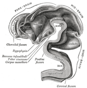
Development
During the first three weeks of
-
 Brain of human embryo at 4.5 weeks, showing interior of forebrain
 -
 Brain interior at 5 weeks
 -
 Brain viewed at midline at 3 months


Function
Cognition
Understanding the
One is obliged to admit that perception and what depends upon it is inexplicable on mechanical principles, that is, by figures and motions. In imagining that there is a machine whose construction would enable it to think, to sense, and to have perception, one could conceive it enlarged while retaining the same proportions, so that one could enter into it, just like into a windmill. Supposing this, one should, when visiting within it, find only parts pushing one another, and never anything by which to explain a perception.
- — Leibniz, Monadology[31]
Doubt about the possibility of a mechanistic explanation of thought drove

Lateralization
Each hemisphere of the brain interacts primarily with one half of the body, but for reasons that are unclear, the connections are crossed: the left side of the brain interacts with the right side of the body, and vice versa.[36] Motor connections from the brain to the spinal cord, and sensory connections from the spinal cord to the brain, both cross the midline at the level of the brainstem. Visual input follows a more complex rule: the optic nerves from the two eyes come together at a point called the optic chiasm, and half of the fibers from each nerve split off to join the other. The result is that connections from the left half of the retina, in both eyes, go to the left side of the brain, whereas connections from the right half of the retina go to the right side of the brain. Because each half of the retina receives light coming from the opposite half of the visual field, the functional consequence is that visual input from the left side of the world goes to the right side of the brain, and vice versa. Thus, the right side of the brain receives somatosensory input from the left side of the body, and visual input from the left side of the visual field—an arrangement that presumably is helpful for visuomotor coordination.
The two cerebral hemispheres are connected by a very large nerve bundle (the largest white matter structure in the brain) called the
In most respects, the left and right sides of the brain are symmetrical in terms of function. For example, the counterpart of the left-hemisphere motor area controlling the right hand is the right-hemisphere area controlling the left hand. There are, however, several very important exceptions, involving language and spatial cognition. In most people, the left hemisphere is "dominant" for language: a stroke that damages a key language area in the left hemisphere can leave the victim unable to speak or understand, whereas equivalent damage to the right hemisphere would cause only minor impairment to language skills.

A substantial part of current understanding of the interactions between the two hemispheres has come from the study of "split-brain patients"—people who underwent surgical transection of the corpus callosum in an attempt to reduce the severity of epileptic seizures. These patients do not show unusual behavior that is immediately obvious, but in some cases can behave almost like two different people in the same body, with the right hand taking an action and then the left hand undoing it. Most of these patients, when briefly shown a picture on the right side of the point of visual fixation, are able to describe it verbally, but when the picture is shown on the left, are unable to describe it, but may be able to give an indication with the left hand of the nature of the object shown.
Language
The study of how language is represented, processed, and acquired by the brain is neurolinguistics, which is a large multidisciplinary field drawing from cognitive neuroscience, cognitive linguistics, and psycholinguistics. This field originated from the 19th-century discovery that damage to different parts of the brain appeared to cause different symptoms: physicians noticed that individuals with damage to a portion of the left inferior frontal gyrus now known as Broca's area had difficulty in producing language (aphasia of speech), whereas those with damage to a region in the left superior temporal gyrus, now known as Wernicke's area, had difficulty in understanding it.[38]

(Associated cortical regions involved in vision, touch sensation, and non-speech movement are also shown.)
Since then, there has been substantial debate over what linguistic processes these and other parts of the brain subserve.[39] Although Broca's and Wernicke's areas have traditionally been associated with language functions, they may also be involved in certain non-speech functions.[citation needed] There is also debate over whether or not there even is a strong one-to-one relationship between brain regions and language functions that emerges during neocortical development.[40] Research on language has increasingly used more modern methods, including electrophysiology and functional neuroimaging, to examine how language processing occurs.
Metabolism

The brain consumes up to twenty percent of the energy used by the human body, more than any other organ.
Although the human brain represents only 2% of the body weight, it receives 15% of the cardiac output, 20% of total body oxygen consumption, and 25% of total body
Clinical significance
Injuries to the brain tend to affect large areas of the organ, sometimes causing major deficits in intelligence, memory, personality, and movement. Head trauma caused, for example, by vehicular or industrial accidents, is a leading cause of death in youth and middle age. In many cases, more damage is caused by resultant edema than by the impact itself. Stroke, caused by the blockage or rupturing of blood vessels in the brain, is another major cause of death from brain damage.
Other problems in the brain can be more accurately classified as diseases.
Some infectious diseases affecting the brain are caused by viruses and bacteria. Infection of the meninges, the membranes that cover the brain, can lead to meningitis. Bovine spongiform encephalopathy (also known as "mad cow disease") is deadly in cattle and humans and is linked to prions. Kuru is a similar prion-borne degenerative brain disease affecting humans, (endemic only to Papua New Guinea tribes). Both are linked to the ingestion of neural tissue, and may explain the tendency in human and some non-human species to avoid cannibalism. Viral or bacterial causes have been reported in multiple sclerosis, and are established causes of encephalopathy, and encephalomyelitis.
Many brain disorders are
Effects of brain damage
A key source of information about the function of brain regions is the effects of damage to them.
Electrodes and magnetic fields
By placing electrodes on the scalp, it is possible to record the summed electrical activity of the cortex using a methodology known as electroencephalography (EEG).[53] EEG records average neuronal activity from the cerebral cortex and can detect changes in activity over large areas but with low sensitivity for sub-cortical activity. EEG recordings are sensitive enough to detect tiny electrical impulses lasting only a few milliseconds. Most EEG devices have good temporal resolution, but low spatial resolution.
Electrodes can also be placed directly on the surface of the brain (usually during surgical procedures that require removal of part of the skull). This technique, called electrocorticography (ECoG), offers finer spatial resolution than electroencephalography, but is very invasive. In addition to measuring the electric field directly via electrodes placed over the skull, it is possible to measure the magnetic field that the brain generates using a method known as magnetoencephalography (MEG).[54] This technique also has good temporal resolution like EEG but with much better spatial resolution. The greatest disadvantage of MEG is that, because the magnetic fields generated by neural activity are very subtle, the neural activity must be relatively close to the surface of the brain to detect its magnetic field. MEGs can only detect the magnetic signatures of neurons located in the depths of cortical folds (sulci) that have dendrites oriented in a way that produces a field.
Imaging
Neuroscientists, along with researchers from allied disciplines, study how the human brain works. Such research has expanded considerably in recent decades. The "Decade of the Brain", an initiative of the United States Government in the 1990s, is considered to have marked much of this increase in research,[55] and was followed in 2013 by the BRAIN Initiative.
Information about the structure and function of the human brain comes from a variety of experimental methods. Most information about the cellular components of the brain and how they work comes from studies of animal subjects, using a variety of techniques. Some techniques, however, are used mainly on humans.
Structural and functional imaging

There are several methods for detecting brain activity changes using three-dimensional imaging of local changes in blood flow. The older methods are
Evolution
The
Studies tend to indicate small to moderate
See also
- Cephalic disorder
- Cephalization
- Common misconceptions about the brain
- Enchanted loom
- Functional specialization (brain)
- History of neuroscience
- Lateralization of brain function
- List of neuroscience databases
- List of regions in the human brain
- Neural development in humans
- Neuroanatomy
- Neuroanthropology
- Outline of the human brain
- Philosophy of mind
- Ten percent of the brain myth
References
- ^ "Cerebrum Etymology". dictionary.com. Retrieved 24 October 2015.
{{cite web}}: Italic or bold markup not allowed in:|publisher=(help) - ^ "Encephalo- Etymology". Online Etymology Dictionary. Retrieved 24 October 2015.
{{cite web}}: Italic or bold markup not allowed in:|publisher=(help) - ^ "Tupaia belangeri". The Genome Institute, Washington University. Retrieved 22 January 2016.
- ISBN 978-0-683-06752-1.
- ^ ISBN 0199920222. Retrieved January 2, 2016.)
{{cite book}}: Cite uses deprecated parameter|authors=(help - PMID 17544382.
- PMID 10234034.
- PMID 19915731.)
there was, to our knowledge, no actual, direct estimate of numbers of cells or of neurons in the entire human brain to be cited until 2009. A reasonable approximation was provided by Williams and Herrup (1988), from the compilation of partial numbers in the literature. These authors estimated the number of neurons in the human brain at about 85 billion [...] With more recent estimates of 21–26 billion neurons in the cerebral cortex (Pelvig et al., 2008 ) and 101 billion neurons in the cerebellum (Andersen et al., 1992 ), however, the total number of neurons in the human brain would increase to over 120 billion neurons.
{{cite journal}}: CS1 maint: unflagged free DOI (link - PMID 19226510.)
despite the widespread quotes that the human brain contains 100 billion neurons and ten times more glial cells, the absolute number of neurons and glial cells in the human brain remains unknown. Here we determine these numbers by using the isotropic fractionator and compare them with the expected values for a human-sized primate. We find that the adult male human brain contains on average 86.1 ± 8.1 billion NeuN-positive cells ("neurons") and 84.6 ± 9.8 billion NeuN-negative ("nonneuronal") cells.
{{cite journal}}: CS1 maint: multiple names: authors list (link - ISBN 978-0-8385-7701-1.
- ISBN 1461448425. Retrieved January 21, 2017.)
{{cite book}}: Cite uses deprecated parameter|authors=(help - ISBN 1136282203. Retrieved January 21, 2017.)
{{cite book}}: Cite uses deprecated parameter|authors=(help - ^ ISBN 049590693X. Retrieved January 21, 2017.
- ISBN 0262019450. Retrieved January 21, 2017.)
{{cite book}}: Cite uses deprecated parameter|authors=(help - ISBN 1107152895. Retrieved January 21, 2017.
- PMID 22992645.
- myelinatedfiber tracts traveling to and from the cerebral cortex.
- ISBN 1461445620. Retrieved January 21, 2017.
- ISBN 1444331213. Retrieved January 21, 2017.
- ISBN 0323101240. Retrieved January 21, 2017.)
{{cite book}}: Cite uses deprecated parameter|authors=(help - PMID 24723857.)
{{cite journal}}: CS1 maint: unflagged free DOI (link - ISSN 0021-9940.
- OCLC 46640860.
- PMID 18267953.
- ^ ISBN 1461452279. Retrieved January 21, 2017.
- ^ ISBN 1118885902. Retrieved January 25, 2017.
- ^ ISBN 0547177798. Retrieved January 25, 2017.
- ^ ISBN 0716795868. Retrieved January 25, 2017.)
{{cite book}}: Cite uses deprecated parameter|authors=(help - ^ Principles of Anatomy and Physiology 12th Edition - Tortora,Page 519.
- ^ Principles of Anatomy and Physiology 12th Edition - Tortora,Page 519-fig. (14.15)
- ISBN 978-0-415-07284-7.
- ^ Hart, WD (1996). Guttenplan S (ed.). A Companion to the Philosophy of Mind. Blackwell. pp. 265–267.
- ISBN 978-0-262-53085-9.
- ^ Aslihan Selimbeyoglu, Josef Parvizi. "Electrical stimulation of the human brain: perceptual and behavioral phenomena reported in the old and new literature" (2010). Frontiers in Human Neuroscience.
- ^ James H. Schwartz. Appendix D: Consciousness and the Neurobiology of the Twenty-First Century. In Kandel, ER; Schwartz JH; Jessell TM. (2000). Principles of Neural Science, 4th Edition.
- ISBN 0470083557. Retrieved January 25, 2017.)
{{cite book}}: Cite uses deprecated parameter|authors=(help - ^ Eric Mooshagian. "Anatomy of the Corpus Callosum Reveals Its Function". Jneurosci.org. Retrieved 2014-03-05.
- ^ Damasio, H. (2001). Neural basis of language disorders. In R. Chapey (Ed.), Language intervention strategies in adult aphasia. 4th edition (pp. 18–36). Baltimore: Williams & Wilkins.
- ^ Regarding the function of Broca's region, see for example the following:
- Grodzinsky, Y (2000). "The neurology of syntax: language use without Broca's area". Behavioral and Brain Sciences. 23 (1): 1–71. .
- Hagoort, P. 2013. MUC (Memory, Unification, Control) and beyond. Frontiers in Language Sciences.
- .
- ^ Swaminathan, Nikhil (29 April 2008). "Why Does the Brain Need So Much Power?". Scientific American. Scientific American, a Division of Nature America, Inc. Retrieved 19 November 2010.
- doi:10.1096/fj.08-106104. Retrieved May 9, 2011.)
{{cite journal}}: CS1 maint: unflagged free DOI (link - PMID 23072752.
- PMID 22403540.)
{{cite journal}}: CS1 maint: unflagged free DOI (link - ISBN 978-0-397-51820-3.)
{{cite book}}: Unknown parameter|editors=ignored (|editor=suggested) (help - PMID 12149485.
- OCLC 44957002.
- ISBN 978-1-4160-6257-8.
- ISBN 978-1-84169-103-9.
- PMID 8969785.
- PMID 19423857.
- PMID 16116107.
- OCLC 43275001.
- OCLC 141379565.
- PMID 10336393. Retrieved 2010-04-05.
- OCLC 45166697.
- ISBN 0787675598. Retrieved January 21, 2017.
As human's position changed and the manner in which his or her skull balanced on the spinal column pivoted, the brain expanded, altering the shape of the cranium.
- ^ "If Modern Humans Are So Smart, Why Are Our Brains Shrinking?". DiscoverMagazine.com. 2011-01-20. Retrieved 2014-03-05.
- .
- PMID 23486442.
- ^ Davidson, Iain. "As large as you need and as small as you can'--implications of the brain size of Homo floresiensis, (Iain Davidson)". Une-au.academia.edu. Retrieved 2011-10-30.
- .
- ^ http://www.uchospitals.edu/news/2005/20050908-humanbrain.html
- )
- PMID 18089578.
- .
- PMID 19323856.
General references
- Campbell, Neil A. and Jane B. Reece. (2005). Biology. Benjamin Cummings. ISBN 0-8053-7171-0
- McGilchrist, Iain (2009). ISBN 0-300-14878-X.
- Ramachanandran, V S (2011), The Tell-Tale Brain: A Neuroscientist's Quest for What Makes Us Human. W. W. Norton & Company.
- Simon, Seymour (1999). The Brain. HarperTrophy. ISBN 0-688-17060-9
- Thompson, Richard F. (2000). The Brain: An Introduction to Neuroscience. Worth Publishers. ISBN 0-7167-3226-2
External links
- Atlas of the Human Brain
- The Whole Brain Atlas
- High-Resolution Cytoarchitectural Primate Brain Atlases
- Brain Facts and Figures
- Interactive Human Brain 3D Tool
| Topics | |
|---|---|
Brain functions | |
| People | |
| Tests |
|
Category:Brain
Brain
Category:Articles with images not understandable by color blind users