
Human brain
| Human brain | |
|---|---|
 The human brain, obtained after an autopsy | |
 Human brain and skull | |
| Details | |
| Precursor | Neural tube |
| System | Central nervous system |
| Artery | Internal carotid arteries, vertebral arteries |
| Vein | Internal jugular vein, internal cerebral veins; external veins: (superior, middle, and inferior cerebral veins), basal vein, and cerebellar veins |
| Identifiers | |
| Latin | encephalon |
| Greek | ἐγκέφαλος (enképhalos)[1] |
| TA98 | A14.1.03.001 |
| TA2 | 5415 |
| FMA | 50801 |
| Anatomical terminology | |
The brain is the central
The cerebrum, the largest part of the human brain, consists of two cerebral hemispheres. Each hemisphere has an inner core composed of white matter, and an outer surface – the cerebral cortex – composed of grey matter. The cortex has an outer layer, the neocortex, and an inner allocortex. The neocortex is made up of six neuronal layers, while the allocortex has three or four. Each hemisphere is divided into four lobes – the frontal, temporal, parietal, and occipital lobes. The frontal lobe is associated with executive functions including self-control, planning, reasoning, and abstract thought, while the occipital lobe is dedicated to vision. Within each lobe, cortical areas are associated with specific functions, such as the sensory, motor, and association regions. Although the left and right hemispheres are broadly similar in shape and function, some functions are associated with one side, such as language in the left and visual-spatial ability in the right. The hemispheres are connected by commissural nerve tracts, the largest being the corpus callosum.
The cerebrum is connected by the brainstem to the spinal cord. The brainstem consists of the
The brain is protected by the
The study of the anatomy of the brain is
In culture, the philosophy of mind has for centuries attempted to address the question of the nature of consciousness and the mind–body problem. The pseudoscience of phrenology attempted to localise personality attributes to regions of the cortex in the 19th century. In science fiction, brain transplants are imagined in tales such as the 1942 Donovan's Brain.
Structure

Gross anatomy
The adult human brain weighs on average about 1.2–1.4 kg (2.6–3.1 lb) which is about 2% of the total body weight,[2][3] with a volume of around 1260 cm3 in men and 1130 cm3 in women.[4] There is substantial individual variation,[4] with the standard reference range for men being 1,180–1,620 g (2.60–3.57 lb)[5] and for women 1,030–1,400 g (2.27–3.09 lb).[6]
The cerebrum, consisting of the cerebral hemispheres, forms the largest part of the brain and overlies the other brain structures.[7] The outer region of the hemispheres, the cerebral cortex, is grey matter, consisting of cortical layers of neurons. Each hemisphere is divided into four main lobes – the frontal lobe, parietal lobe, temporal lobe, and occipital lobe.[8] Three other lobes are included by some sources which are a central lobe, a limbic lobe, and an insular lobe.[9] The central lobe comprises the precentral gyrus and the postcentral gyrus and is included since it forms a distinct functional role.[9][10]
The
The cerebrum, brainstem, cerebellum, and spinal cord are covered by four[11] membranes called meninges. The membranes are the tough dura mater; the middle arachnoid mater and the more delicate inner pia mater. Between the arachnoid mater and the pia mater is the subarachnoid space and subarachnoid cisterns, which contain the cerebrospinal fluid.[12] The outermost membrane of the cerebral cortex is the basement membrane of the pia mater called the glia limitans and is an important part of the blood–brain barrier.[13] The living brain is very soft, having a gel-like consistency similar to soft tofu.[14] The cortical layers of neurons constitute much of the cerebral grey matter, while the deeper subcortical regions of myelinated axons, make up the white matter.[7] The white matter of the brain makes up about half of the total brain volume.[15]

.png)
Cerebrum


The cerebrum is the largest part of the brain and is divided into nearly symmetrical left and right hemispheres by a deep groove, the longitudinal fissure.[16] Asymmetry between the lobes is noted as a petalia.[17] The hemispheres are connected by five commissures that span the longitudinal fissure, the largest of these is the corpus callosum.[7] Each hemisphere is conventionally divided into four main
The outer part of the cerebrum is the cerebral cortex, made up of grey matter arranged in layers. It is 2 to 4 millimetres (0.079 to 0.157 in) thick, and deeply folded to give a convoluted appearance.[20] Beneath the cortex is the cerebral white matter. The largest part of the cerebral cortex is the neocortex, which has six neuronal layers. The rest of the cortex is of allocortex, which has three or four layers.[7]
The cortex is

The cerebrum contains the ventricles where the cerebrospinal fluid is produced and circulated. Below the corpus callosum is the septum pellucidum, a membrane that separates the lateral ventricles. Beneath the lateral ventricles is the thalamus and to the front and below is the hypothalamus. The hypothalamus leads on to the pituitary gland. At the back of the thalamus is the brainstem.[26]
The basal ganglia, also called basal nuclei, are a set of structures deep within the hemispheres involved in behaviour and movement regulation.[27] The largest component is the striatum, others are the globus pallidus, the substantia nigra and the subthalamic nucleus.[27] The striatum is divided into a ventral striatum, and dorsal striatum, subdivisions that are based upon function and connections. The ventral striatum consists of the nucleus accumbens and the olfactory tubercle whereas the dorsal striatum consists of the caudate nucleus and the putamen. The putamen and the globus pallidus lie separated from the lateral ventricles and thalamus by the internal capsule, whereas the caudate nucleus stretches around and abuts the lateral ventricles on their outer sides.[28] At the deepest part of the lateral sulcus between the insular cortex and the striatum is a thin neuronal sheet called the claustrum.[29]
Below and in front of the striatum are a number of basal forebrain structures. These include the nucleus basalis, diagonal band of Broca, substantia innominata, and the medial septal nucleus. These structures are important in producing the neurotransmitter, acetylcholine, which is then distributed widely throughout the brain. The basal forebrain, in particular the nucleus basalis, is considered to be the major cholinergic output of the central nervous system to the striatum and neocortex.[30]
Cerebellum

The cerebellum is divided into an anterior lobe, a posterior lobe, and the flocculonodular lobe.[31] The anterior and posterior lobes are connected in the middle by the vermis.[32] Compared to the cerebral cortex, the cerebellum has a much thinner outer cortex that is narrowly furrowed into numerous curved transverse fissures.[32] Viewed from underneath between the two lobes is the third lobe the flocculonodular lobe.[33] The cerebellum rests at the back of the cranial cavity, lying beneath the occipital lobes, and is separated from these by the cerebellar tentorium, a sheet of fibre.[34]
It is connected to the brainstem by three pairs of
Brainstem
The brainstem lies beneath the cerebrum and consists of the
Ten of the twelve pairs of
Microanatomy
The human brain is primarily composed of
Types of glial cell are
Some 400
Cerebrospinal fluid

Cerebrospinal fluid is a clear, colourless
A
Blood supply


The
The internal carotid arteries are branches of the
The vertebral arteries emerge as branches of the left and right
Blood drainage
Cerebral veins drain deoxygenated blood from the brain. The brain has two main networks of veins: an exterior or superficial network, on the surface of the cerebrum that has three branches, and an interior network. These two networks communicate via anastomosing (joining) veins.[60] The veins of the brain drain into larger cavities of the dural venous sinuses usually situated between the dura mater and the covering of the skull.[61] Blood from the cerebellum and midbrain drains into the great cerebral vein. Blood from the medulla and pons of the brainstem have a variable pattern of drainage, either into the spinal veins or into adjacent cerebral veins.[60]
The blood in the deep part of the brain drains, through a venous plexus into the cavernous sinus at the front, and the superior and inferior petrosal sinuses at the sides, and the inferior sagittal sinus at the back.[61] Blood drains from the outer brain into the large superior sagittal sinus, which rests in the midline on top of the brain. Blood from here joins with blood from the straight sinus at the confluence of sinuses.[61]
Blood from here drains into the left and right
The blood–brain barrier
The larger arteries throughout the brain supply blood to smaller
Development


At the beginning of the third week of development, the embryonic ectoderm forms a thickened strip called the neural plate.[64] By the fourth week of development the neural plate has widened to give a broad cephalic end, a less broad middle part and a narrow caudal end. These swellings are known as the primary brain vesicles and represent the beginnings of the forebrain (prosencephalon), midbrain (mesencephalon), and hindbrain (rhombencephalon).[65][66]
Neural crest cells (derived from the ectoderm) populate the lateral edges of the plate at the neural folds. In the fourth week—during the neurulation stage—the neural folds close to form the neural tube, bringing together the neural crest cells at the neural crest.[67] The neural crest runs the length of the tube with cranial neural crest cells at the cephalic end and caudal neural crest cells at the tail. Cells detach from the crest and migrate in a craniocaudal (head to tail) wave inside the tube.[67] Cells at the cephalic end give rise to the brain, and cells at the caudal end give rise to the spinal cord.[68]
The tube flexes as it grows, forming the crescent-shaped cerebral hemispheres at the head. The cerebral hemispheres first appear on day 32.[69] Early in the fourth week, the cephalic part bends sharply forward in a
A characteristic of the brain is the cortical folding known as gyrification. For just over five months of prenatal development the cortex is smooth. By the gestational age of 24 weeks, the wrinkled morphology showing the fissures that begin to mark out the lobes of the brain is evident.[74] Why the cortex wrinkles and folds is not well-understood, but gyrification has been linked to intelligence and neurological disorders, and a number of gyrification theories have been proposed.[74] These theories include those based on mechanical buckling,[75][18] axonal tension,[76] and differential tangential expansion.[75] What is clear is that gyrification is not a random process, but rather a complex developmentally predetermined process which generates patterns of folds that are consistent between individuals and most species.[75][77]
The first groove to appear in the fourth month is the lateral cerebral fossa.[69] The expanding caudal end of the hemisphere has to curve over in a forward direction to fit into the restricted space. This covers the fossa and turns it into a much deeper ridge known as the lateral sulcus and this marks out the temporal lobe.[69] By the sixth month other sulci have formed that demarcate the frontal, parietal, and occipital lobes.[69] A gene present in the human genome (ARHGAP11B) may play a major role in gyrification and encephalisation.[78]
-
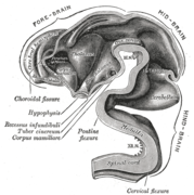
 Brain of human embryo at 4.5 weeks, showing interior of forebrain
 -
 Brain interior at 5 weeks
 -
 Brain viewed at midline at 3 months


Function

Motor control
The frontal lobe is involved in reasoning, motor control, emotion, and language. It contains the
Gross movement – such as
Sensory


The
From the skin, the brain receives information about
The sense of
Regulation
Autonomic functions of the brain include the regulation, or rhythmic control of the heart rate and rate of breathing, and maintaining homeostasis.
The brain controls the
The
The
Language

While language functions were traditionally thought to be localised to Wernicke's area and Broca's area,[102] it is now mostly accepted that a wider network of cortical regions contributes to language functions.[103][104][105]
The study on how language is represented, processed, and acquired by the brain is called neurolinguistics, which is a large multidisciplinary field drawing from cognitive neuroscience, cognitive linguistics, and psycholinguistics.[106]
Lateralisation
The cerebrum has a
The left and right sides of the brain appear symmetrical, but they function asymmetrically.[113] For example, the counterpart of the left-hemisphere motor area controlling the right hand is the right-hemisphere area controlling the left hand. There are, however, several important exceptions, involving language and spatial cognition. The left frontal lobe is dominant for language. If a key language area in the left hemisphere is damaged, it can leave the victim unable to speak or understand,[113] whereas equivalent damage to the right hemisphere would cause only minor impairment to language skills.
A substantial part of current understanding of the interactions between the two hemispheres has come from the study of "split-brain patients"—people who underwent surgical transection of the corpus callosum in an attempt to reduce the severity of epileptic seizures.[114] These patients do not show unusual behaviour that is immediately obvious, but in some cases can behave almost like two different people in the same body, with the right hand taking an action and then the left hand undoing it.[114][115] These patients, when briefly shown a picture on the right side of the point of visual fixation, are able to describe it verbally, but when the picture is shown on the left, are unable to describe it, but may be able to give an indication with the left hand of the nature of the object shown.[115][116]
Emotion
Cognition
The brain is responsible for
The
Physiology
Neurotransmission
Brain activity is made possible by the interconnections of
Metabolism

The brain consumes up to 20% of the energy used by the human body, more than any other organ.
Although the human brain represents only 2% of the body weight, it receives 15% of the cardiac output, 20% of total body oxygen consumption, and 25% of total body
The function of
Research
The brain is not fully understood, and research is ongoing.[146] Neuroscientists, along with researchers from allied disciplines, study how the human brain works. The boundaries between the specialties of neuroscience, neurology and other disciplines such as psychiatry have faded as they are all influenced by basic research in neuroscience.
Neuroscience research has expanded considerably. The "Decade of the Brain", an initiative of the United States Government in the 1990s, is considered to have marked much of this increase in research,[147] and was followed in 2013 by the BRAIN Initiative.[148] The Human Connectome Project was a five-year study launched in 2009 to analyse the anatomical and functional connections of parts of the brain, and has provided much data.[146]
An emerging phase in research may be that of simulating brain activity.[149]
Methods
Information about the structure and function of the human brain comes from a variety of experimental methods, including animals and humans. Information about brain trauma and stroke has provided information about the function of parts of the brain and the effects of
Invasive measures include electrocorticography, which uses electrodes placed directly on the exposed surface of the brain. This method is used in cortical stimulation mapping, used in the study of the relationship between cortical areas and their systemic function.[152] By using much smaller microelectrodes, single-unit recordings can be made from a single neuron that give a high spatial resolution and high temporal resolution. This has enabled the linking of brain activity to behaviour, and the creation of neuronal maps.[153]
The development of cerebral organoids has opened ways for studying the growth of the brain, and of the cortex, and for understanding disease development, offering further implications for therapeutic applications.[154][155]
Imaging
Any electrical current generates a magnetic field;
Differences in
Advances in neuroimaging have enabled objective insights into mental disorders, leading to faster diagnosis, more accurate prognosis, and better monitoring.[162]
Gene and protein expression
Bioinformatics is a field of study that includes the creation and advancement of databases, and computational and statistical techniques, that can be used in studies of the human brain, particularly in the areas of gene and protein expression. Bioinformatics and studies in genomics, and functional genomics, generated the need for DNA annotation, a transcriptome technology, identifying genes, their locations and functions.[163][164][165] GeneCards is a major database.
As of 2017[update], just under 20,000 protein-coding genes are seen to be expressed in the human,[163] and some 400 of these genes are brain-specific.[166][167] The data that has been provided on gene expression in the brain has fuelled further research into a number of disorders. The long term use of alcohol for example, has shown altered gene expression in the brain, and cell-type specific changes that may relate to alcohol use disorder.[168] These changes have been noted in the synaptic transcriptome in the prefrontal cortex, and are seen as a factor causing the drive to alcohol dependence, and also to other substance abuses.[169]
Other related studies have also shown evidence of synaptic alterations and their loss, in the
Clinical significance
Injury
Disease
Cerebral atherosclerosis is atherosclerosis that affects the brain. It results from the build-up of plaques formed of cholesterol, in the large arteries of the brain, and can be mild to significant. When significant, arteries can become narrowed enough to reduce blood flow. It contributes to the development of dementia, and has protein similarities to those found in Alzheimer’s disease.[175]
The brain, although protected by the blood–brain barrier, can be affected by infections including
Tumours
Mental disorders
Epilepsy
Congenital
Some brain disorders, such as
Most cerebral arteriovenous malformations are congenital, these tangled networks of blood vessels may remain without symptoms but at their worst may rupture and cause intracranial hemorrhaging.[189]Stroke
A
Most strokes result from loss of blood supply, typically because of an
Some treatments for stroke are time-critical. These include
Having experienced a stroke, a person may be admitted to a
Brain death
Brain death refers to an irreversible total loss of brain function.
When brain death is suspected, reversible
Society and culture
Neuroanthropology is the study of the relationship between culture and the brain. It explores how the brain gives rise to culture, and how culture influences brain development.[206] Cultural differences and their relation to brain development and structure are researched in different fields.[207]
The mind

The
One is obliged to admit that perception and what depends upon it is inexplicable on mechanical principles, that is, by figures and motions. In imagining that there is a machine whose construction would enable it to think, to sense, and to have perception, one could conceive it enlarged while retaining the same proportions, so that one could enter into it, just like into a windmill. Supposing this, one should, when visiting within it, find only parts pushing one another, and never anything by which to explain a perception.
- — Leibniz, Monadology[209]
Doubt about the possibility of a mechanistic explanation of thought drove
Brain size
The size of the brain and a person's intelligence are not strongly related.[214] Studies tend to indicate small to moderate correlations (averaging around 0.3 to 0.4) between brain volume and IQ.[215] The most consistent associations are observed within the frontal, temporal, and parietal lobes, the hippocampi, and the cerebellum, but these only account for a relatively small amount of variance in IQ, which itself has only a partial relationship to general intelligence and real-world performance.[216][217]
Other animals, including whales and elephants have larger brains than humans. However, when the
In popular culture

Earlier ideas about the relative importance of the different organs of the human body sometimes emphasised the heart.[219] Modern Western popular conceptions, in contrast, have placed increasing focus on the
Research has disproved some common misconceptions about the brain. These include both ancient and modern myths. It is not true (for example) that neurons are not replaced after the age of two; nor that normal humans use only ten per cent of the brain.[221] Popular culture has also oversimplified the lateralisation of the brain by suggesting that functions are completely specific to one side of the brain or the other. Akio Mori coined the term "game brain" for the unreliably supported theory that spending long periods playing video games harmed the brain's pre-frontal region, and impaired the expression of emotion and creativity.[222]
Historically, particularly in the early-19th century, the brain featured in popular culture through phrenology, a pseudoscience that assigned personality attributes to different regions of the cortex. The cortex remains important in popular culture as covered in books and satire.[223][224]
The human brain can feature in
History
Early history
The
In the fifth century BC,
Renaissance

In 1316, Mondino de Luzzi's Anathomia began the modern study of brain anatomy.[231]
In the middle of 19th century Emil du Bois-Reymond and Hermann von Helmholtz were able to use a galvanometer to show that electrical impulses passed at measurable speeds along nerves, refuting the view of their teacher Johannes Peter Müller that the nerve impulse was a vital function that could not be measured.[239][240][241] Richard Caton in 1875 demonstrated electrical impulses in the cerebral hemispheres of rabbits and monkeys.[242] In the 1820s, Jean Pierre Flourens pioneered the experimental method of damaging specific parts of animal brains describing the effects on movement and behavior.[243]
Modern period


Studies of the brain became more sophisticated with the use of the
Charles Sherrington published his influential 1906 work The Integrative Action of the Nervous System examining the function of reflexes, evolutionary development of the nervous system, functional specialisation of the brain, and layout and cellular function of the central nervous system.[245] In 1942 he coined the term enchanted loom as a metaphor for the brain. John Farquhar Fulton, founded the Journal of Neurophysiology and published the first comprehensive textbook on the physiology of the nervous system during 1938.[246] Neuroscience during the twentieth century began to be recognised as a distinct unified academic discipline, with David Rioch, Francis O. Schmitt, and Stephen Kuffler playing critical roles in establishing the field.[247] Rioch originated the integration of basic anatomical and physiological research with clinical psychiatry at the Walter Reed Army Institute of Research, starting in the 1950s.[248] During the same period, Schmitt established the Neuroscience Research Program, an inter-university and international organisation, bringing together biology, medicine, psychological and behavioural sciences. The word neuroscience itself arises from this program.[249]
Harvey Cushing (1869–1939) is recognised as the first proficient brain surgeon in the world.[254] In 1937, Walter Dandy began the practice of vascular neurosurgery by performing the first surgical clipping of an intracranial aneurysm.[255]
Comparative anatomy
The human brain has many properties that are common to all vertebrate brains.[256] Many of its features are common to all mammalian brains,[257] most notably a six-layered cerebral cortex and a set of associated structures,[258] including the hippocampus and amygdala.[259] The cortex is proportionally larger in humans than in many other mammals.[260] Humans have more association cortex, sensory and motor parts than smaller mammals such as the rat and the cat.[261]
As a primate brain, the human brain has a much larger cerebral cortex, in proportion to body size, than most mammals,[259] and a highly developed visual system.[262][263]
As a
See also
- Outline of the human brain
- Outline of neuroscience
- Cerebral atrophy
- Cortical spreading depression
- Evolution of human intelligence
- Large-scale brain networks
- Superficial veins of the brain
References
- ^ "Encephalo- Etymology". Online Etymology Dictionary. Archived from the original on October 2, 2017. Retrieved October 24, 2015.
- ISBN 978-0-683-06752-1.
- ^ ISBN 978-0-19-992022-8.
- ^ PMID 17544382.
- S2CID 32174574.
- S2CID 25319215.
- ^ a b c d e Gray's Anatomy 2008, pp. 227–9.
- ^ a b Gray's Anatomy 2008, pp. 335–7.
- ^ PMID 20121437.
- PMID 25555079.
- S2CID 255440992.
- University of Rochester Medical Center (January 5, 2023). "Newly discovered anatomy shields and monitors brain". Medical Xpress. Archived from the original on January 7, 2023. Retrieved February 15, 2023.
- ^ Purves 2012, p. 724.
- ^ a b Cipolla, M.J. (January 1, 2009). "Anatomy and Ultrastructure". The Cerebral Circulation. Morgan & Claypool Life Sciences. Archived from the original on October 1, 2017 – via NCBI Bookshelf.
- ^ "A Surgeon's-Eye View of the Brain". NPR. Fresh Air. May 10, 2006. Archived from the original on November 7, 2017.
- PMID 29268094.
- ISBN 978-1-4443-3121-9.
- S2CID 58546404.
- ^ ISBN 978-0-309-04529-2.
- ^ Larsen 2001, pp. 455–456.
- ISBN 978-0-8385-7701-1.
- ^ Guyton & Hall 2011, p. 574.
- ^ Guyton & Hall 2011, p. 667.
- ^ Principles of Anatomy and Physiology 12th Edition – Tortora, p. 519.
- ^ ISBN 978-0-547-17779-3.
- ^ ISBN 978-0-7167-9586-5.
- ^ Pocock 2006, p. 64.
- ^ a b Purves 2012, p. 399.
- ^ Gray's Anatomy 2008, pp. 325–6.
- S2CID 38353825.
- PMID 19801988.
- ^ Guyton & Hall 2011, p. 699.
- ^ a b c Gray's Anatomy 2008, p. 298.
- ISBN 978-1-4557-0418-7.
- ^ a b Gray's Anatomy 2008, p. 297.
- ^ Guyton & Hall 2011, pp. 698–9.
- ^ Squire 2013, pp. 761–763.
- ^ a b c d e f Gray's Anatomy 2008, p. 275.
- ^ Guyton & Hall 2011, p. 691.
- ^ Purves 2012, p. 377.
- ^ S2CID 5200449.
despite the widespread quotes that the human brain contains 100 billion neurons and ten times more glial cells, the absolute number of neurons and glial cells in the human brain remains unknown. Here we determine these numbers by using the isotropic fractionator and compare them with the expected values for a human-sized primate. We find that the adult male human brain contains on average 86.1 ± 8.1 billion NeuN-positive cells ("neurons") and 84.6 ± 9.8 billion NeuN-negative ("nonneuronal") cells.
- ISBN 978-80-246-2067-1.
- ^ PMID 26377554.
- ^ a b c d e Guyton & Hall 2011, pp. 748–749.
- PMID 24833851.
- PMID 25830558.
- PMID 26076492.
- ^ a b c d Gray's Anatomy 2008, pp. 242–244.
- ^ Purves 2012, p. 742.
- ^ Gray's Anatomy 2008, p. 243.
- PMID 23709744.
- ^ Gaillard, F. "Glymphatic pathway". radiopaedia.org. Archived from the original on October 30, 2017.
- ^ PMID 29163074.
The paravascular pathway, also known as the "glymphatic" pathway, is a recently described system for waste clearance in the brain. According to this model, cerebrospinal fluid (CSF) enters the paravascular spaces surrounding penetrating arteries of the brain, mixes with interstitial fluid (ISF) and solutes in the parenchyma, and exits along paravascular spaces of draining veins. ... In addition to Aβ clearance, the glymphatic system may be involved in the removal of other interstitial solutes and metabolites. By measuring the lactate concentration in the brains and cervical lymph nodes of awake and sleeping mice, Lundgaard et al. (2017) demonstrated that lactate can exit the CNS via the paravascular pathway. Their analysis took advantage of the substantiated hypothesis that glymphatic function is promoted during sleep (Xie et al., 2013; Lee et al., 2015; Liu et al., 2017).
- PMID 26425672.
- ^ S2CID 6290040.
- ^ Gray's Anatomy 2008, p. 247.
- ^ Gray's Anatomy 2008, pp. 251–2.
- ^ a b c Gray's Anatomy 2008, p. 250.
- ^ a b Gray's Anatomy 2008, p. 248.
- ^ a b Gray's Anatomy 2008, p. 251.
- ^ a b c Gray's Anatomy 2008, pp. 254–6.
- ^ a b c d e Elsevier's 2007, pp. 311–4.
- PMID 20944625.
- ^ Laterra, J.; Keep, R.; Betz, L.A.; et al. (1999). "Blood–cerebrospinal fluid barrier". Basic neurochemistry: molecular, cellular and medical aspects (6th ed.). Philadelphia: Lippincott-Raven.
- ISBN 978-0-7817-9069-7.
- ^ a b Larsen 2001, p. 419.
- S2CID 260286574.
- ^ a b c Larsen 2001, pp. 85–88.
- ^ Purves 2012, pp. 480–482.
- ^ a b c d Larsen 2001, pp. 445–446.
- ^ "OpenStax CNX". cnx.org. Archived from the original on May 5, 2015. Retrieved May 5, 2015.
- ^ Larsen 2001, pp. 85–87.
- ^ Purves 2012, pp. 481–484.
- ISBN 978-0-87893-742-4.
- ^ ISBN 978-1-4614-4562-3.
- ^ PMID 23542881.
- S2CID 4355025.
- PMID 29367288.
- S2CID 34506325.
- ^ "Parts of the Brain | Introduction to Psychology". courses.lumenlearning.com. Retrieved September 20, 2019.
- ^ Guyton & Hall 2011, p. 685.
- ^ a b Guyton & Hall 2011, p. 687.
- ^ a b Guyton & Hall 2011, p. 686.
- ^ Guyton & Hall 2011, pp. 698, 708.
- ^ Davidson's 2010, p. 1139.
- ^ ISBN 978-1-61069-338-7.
- ^ a b Guyton & Hall 2011, pp. 571–576.
- ^ Guyton & Hall 2011, pp. 573–574.
- ^ Guyton & Hall 2011, pp. 623–631.
- ^ Guyton & Hall 2011, pp. 739–740.
- ^ Pocock 2006, pp. 138–139.
- ^ Squire 2013, pp. 525–526.
- ^ Guyton & Hall 2011, pp. 647–648.
- ^ Guyton & Hall 2011, pp. 202–203.
- ^ Guyton & Hall 2011, pp. 205–208.
- ^ a b c d Guyton & Hall 2011, pp. 505–509.
- ^ "Brain Basics: Understanding Sleep | National Institute of Neurological Disorders and Stroke". www.ninds.nih.gov. Archived from the original on December 22, 2017.
- ^ Guyton & Hall 2011, p. 723.
- ISBN 978-0-387-92271-3.
- ^ Squire 2013, p. 800.
- ^ Squire 2013, p. 803.
- ^ Squire 2013, p. 805.
- ^ Guyton & Hall 2011, pp. 720–2.
- PMID 23055482.
- PMID 20161054.
- S2CID 2833893.
- OCLC 45952164.
- S2CID 7399128.
- ISBN 978-1-61069-338-7.
- ISBN 978-1-4641-3960-4.
- ISBN 978-0-470-08355-0.
- ISBN 978-1-133-70853-7.
- ISBN 978-1-305-46529-9.
- ^ ISBN 978-0-387-49985-7.
- ^ ISBN 978-0-205-76906-3.
- ^ ISBN 978-1-4641-3960-4.
- ISBN 978-1-4292-1821-4.
- ISBN 978-0-521-17155-7.
- PMID 22617651.
- S2CID 7150871.
- ISBN 978-0-07-148127-4.
- ^ ISBN 978-0-07-182770-6.
- ^ ISBN 978-0-07-182770-6.
- ^ PMID 23020641.
Figure 4: Executive functions and related terms Archived May 9, 2018, at the Wayback Machine - ^ ISBN 978-1-4614-8106-5.
- ^ ISBN 978-0-07-182770-6.
In conditions in which prepotent responses tend to dominate behavior, such as in drug addiction, where drug cues can elicit drug seeking (Chapter 16), or in attention deficit hyperactivity disorder (ADHD; described below), significant negative consequences can result. ... ADHD can be conceptualized as a disorder of executive function; specifically, ADHD is characterized by reduced ability to exert and maintain cognitive control of behavior. Compared with healthy individuals, those with ADHD have diminished ability to suppress inappropriate prepotent responses to stimuli (impaired response inhibition) and diminished ability to inhibit responses to irrelevant stimuli (impaired interference suppression). ... Functional neuroimaging in humans demonstrates activation of the prefrontal cortex and caudate nucleus (part of the dorsal striatum) in tasks that demand inhibitory control of behavior. ... Early results with structural MRI show a thinner cerebral cortex, across much of the cerebrum, in ADHD subjects compared with age-matched controls, including areas of [the] prefrontal cortex involved in working memory and attention.
- ^ Pocock 2006, p. 68.
- PMID 20007821.
- ^ Pocock 2006, pp. 70–74.
- ^ a b "NIMH » Brain Basics". www.nimh.nih.gov. Archived from the original on March 26, 2017. Retrieved March 26, 2017.
- ISBN 978-0-87893-695-3.
- ^ Swaminathan, N (April 29, 2008). "Why Does the Brain Need So Much Power?". Scientific American. Archived from the original on January 27, 2014. Retrieved November 19, 2010.
- ^ PMID 18840763.
Four grams of glucose circulates in the blood of a person weighing 70 kg. This glucose is critical for normal function in many cell types. In accordance with the importance of these 4 g of glucose, a sophisticated control system is in place to maintain blood glucose constant. Our focus has been on the mechanisms by which the flux of glucose from liver to blood and from blood to skeletal muscle is regulated. ... The brain consumes ~60% of the blood glucose used in the sedentary, fasted person. ... The amount of glucose in the blood is preserved at the expense of glycogen reservoirs (Fig. 2). In postabsorptive humans, there are ~100 g of glycogen in the liver and ~400 g of glycogen in muscle. Carbohydrate oxidation by the working muscle can go up by ~10-fold with exercise, and yet after 1 h, blood glucose is maintained at ~4 g. ... It is now well established that both insulin and exercise cause translocation of GLUT4 to the plasma membrane. Except for the fundamental process of GLUT4 translocation, [muscle glucose uptake (MGU)] is controlled differently with exercise and insulin. Contraction-stimulated intracellular signaling (52, 80) and MGU (34, 75, 77, 88, 91, 98) are insulin independent. Moreover, the fate of glucose extracted from the blood is different in response to exercise and insulin (91, 105). For these reasons, barriers to glucose flux from blood to muscle must be defined independently for these two controllers of MGU.
- S2CID 15394163.
- PMID 22403540.
- PMID 23072752.
- PMID 15717057.
Uptake of valproic acid was reduced in the presence of medium-chain fatty acids such as hexanoate, octanoate, and decanoate, but not propionate or butyrate, indicating that valproic acid is taken up into the brain via a transport system for medium-chain fatty acids, not short-chain fatty acids. ... Based on these reports, valproic acid is thought to be transported bidirectionally between blood and brain across the BBB via two distinct mechanisms, monocarboxylic acid-sensitive and medium-chain fatty acid-sensitive transporters, for efflux and uptake, respectively.
- PMID 23789956.
Monocarboxylate transporters (MCTs) are known to mediate the transport of short chain monocarboxylates such as lactate, pyruvate and butyrate. ... MCT1 and MCT4 have also been associated with the transport of short chain fatty acids such as acetate and formate which are then metabolized in the astrocytes [78].
- ISBN 978-0-397-51820-3.
- ISBN 978-1-4684-3348-7.
- PMID 12149485.
- ISBN 978-0-387-09488-5.
- PMID 25157149.
- ^ "Brain may flush out toxins during sleep". National Institutes of Health. Archived from the original on October 20, 2013. Retrieved October 25, 2013.
- PMID 24136970.
Thus, the restorative function of sleep may be a consequence of the enhanced removal of potentially neurotoxic waste products that accumulate in the awake central nervous system.
- S2CID 54052089. Archived from the original(PDF) on December 26, 2018.
- ^ PMID 22366334.
- S2CID 13261978.
- ^ "A $4.5 Billion Price Tag for the BRAIN Initiative?". Science | AAAS. June 5, 2014. Archived from the original on June 18, 2017.
- PMID 31133838.
- PMID 7678386.
- ^ Purves 2012, pp. 632–633.
- from the original on November 17, 2012.
- S2CID 23389986.
- PMID 23995685.
- PMID 28822354.
- ^ "Magnetic Resonance, a critical peer-reviewed introduction; functional MRI". European Magnetic Resonance Forum. Archived from the original on June 2, 2017. Retrieved June 30, 2017.
- S2CID 8736954.
- S2CID 93823.
- ^ Purves 2012, p. 20.
- ISBN 978-0-19-023473-7.
Irimia, Chambers, Torgerson, and Van Horn (2012) provide a first-step graphic on how best to display connectivity findings, as is presented in Figure 13.15. This is referred to as a connectogram.
- ISBN 978-1-84169-103-9.
- ^ Lepage, M. (2010). "Research at the Brain Imaging Centre". Douglas Mental Health University Institute. Archived from the original on March 5, 2012.
- ^ PMID 28558813.
- PMID 22955987.
- ISBN 9780878932368.
- ^ "The human proteome in brain – The Human Protein Atlas". www.proteinatlas.org. Archived from the original on September 29, 2017. Retrieved September 29, 2017.
- S2CID 802377.
- PMID 28254370.
- PMID 25896098.
- PMID 28497576.
- PMID 25386117.
- ^ "Brain Injury, Traumatic". Medcyclopaedia. GE. Archived from the original on May 26, 2011.
- ^ Dawodu, S.T. (March 9, 2017). "Traumatic Brain Injury (TBI) – Definition and Pathophysiology: Overview, Epidemiology, Primary Injury". Medscape. Archived from the original on April 9, 2017.
- ^ Davidson's 2010, pp. 1196–7.
- PMID 32424284.
- ^ a b Davidson's 2010, pp. 1205–15.
- ^ a b c d e Davidson's 2010, pp. 1216–7.
- PMID 26816013.
- ISBN 978-0-231-53609-7.
- ^ a b c d Davidson's 2010, pp. 1172–9.
- ^ "Status Epilepticus". Epilepsy Foundation.
- ISBN 978-1-4051-0459-3.
- ISBN 978-1-60623-786-1.
- ISBN 978-1-898683-56-8.
- ^ ISBN 978-0-8036-2999-8.
- ISBN 978-1-337-09810-6.
- ISBN 978-0-521-80691-6.
- ISBN 978-0-470-65457-6.
- ^ "Arteriovenous Malformations (AVMs) | National Institute of Neurological Disorders and Stroke". www.ninds.nih.gov. Retrieved February 8, 2023.
- S2CID 36692451.
- ^ Davidson's 2010, p. 1183.
- ^ a b Davidson's 2010, pp. 1180–1.
- ^ Davidson's 2010, pp. 1181, 1183–1185.
- ^ a b c d e f Davidson's 2010, pp. 1183–1185.
- ^ a b Davidson's 2010, pp. 1185–1189.
- S2CID 34799180.
- PMID 16339467.
- S2CID 4967333.
- PMID 30177276.
- ^ PMID 19881172.
- ^ S2CID 219203458.
- PMID 25249744.
- ^ a b c d Davidson's 2010, p. 1158.
- ^ Davidson's 2010, p. 200.
- ISBN 978-0-323-29414-0.
- )
- ^ "Cultural Environment Influences Brain Function | Psych Central News". Psych Central News. August 4, 2010. Archived from the original on January 17, 2017.
- ^ ISBN 978-0-262-13363-0.
- ISBN 978-0-415-07284-7.
- ^ Hart, WD (1996). Guttenplan S (ed.). A Companion to the Philosophy of Mind. Blackwell. pp. 265–267.
- ISBN 978-0-262-53085-9.
- PMID 20577584.
- ^ Schwartz, J.H. Appendix D: Consciousness and the Neurobiology of the Twenty-First Century. In Kandel, E.R.; Schwartz, J.H.; Jessell, T.M. (2000). Principles of Neural Science, 4th Edition.
- ISBN 978-1-4443-6074-5.
- (PDF) from the original on September 6, 2014.
- PMID 18089578.
- .
- ^ "Tupaia belangeri". The Genome Institute, Washington University. Archived from the original on June 1, 2010. Retrieved January 22, 2016.
- ^
Carrier, Martin; ISBN 9783110128765. Retrieved May 22, 2021.
[...] the Aristotelian view that the soul resides primarily in the heart [...].
- ^
ISBN 9781541646865. Retrieved May 22, 2021.
[...] the ways in which we think about [the brain] are much richer than in the past, not simply because of the amazing facts we have discovered, but above all because of how we interpret them.
- ISBN 978-1-118-31271-1.
- ^ Phillips, Helen (July 11, 2002). "Video game "brain damage" claim criticised". New Scientist. Archived from the original on January 11, 2009. Retrieved February 6, 2008.
- ^ Popova, Maria (August 18, 2011). "'Brain Culture': How Neuroscience Became a Pop Culture Fixation". The Atlantic. Archived from the original on July 28, 2017.
- ISBN 978-0-8135-5013-8.
- ^ Cyborgs and Space Archived October 6, 2011, at the Wayback Machine, in Astronautics (September 1960), by Manfred E. Clynes and Nathan S. Kline.
- ISBN 978-1-57181-538-5.
- ISBN 978-0-8385-7701-1.
- ^ ISBN 978-0-8176-3335-6. Archived(PDF) from the original on May 5, 2013.
- ^ ISBN 978-0-7817-3944-3.
- ^ von Staden, p.157
- ISBN 978-0-19-534062-4.
- ^ a b Lokhorst, Gert-Jan (January 1, 2016). "Descartes and the Pineal Gland". The Stanford Encyclopedia of Philosophy. Metaphysics Research Lab, Stanford University. Retrieved March 11, 2017.
- ^ ISBN 978-0-262-57135-7.
- ISBN 978-1-475-74997-7.
- ISBN 978-0-19-970681-5.
- PMID 12470188.
- PMID 12612118.
- PMC 1468164.
- ^ Olesko, Kathryn M.; Holmes, Frederic L. (1994). Cahan, David (ed.). "Experiment, Quantification, and Discovery: Helmholtz's Early Physiological Researches, 1843-50". Hermann von Helmholtz and the Foundations of Nineteenth Century Science. Berkeley; Los Angeles; London: University of California Press: 50–108.
{{cite journal}}: Cite journal requires|journal=(help) - ^ Sabbatini, Renato M.E. "Sabbatini, R.M.E.: The Discovery of Bioelectricity. Nerve Conduction". www.cerebromente.org.br. Archived from the original on June 26, 2017. Retrieved June 10, 2017.
- )
- PMID 2192889.
- PMID 19295220.
- ^ S2CID 7266966.
- PMID 17438014.
- ISBN 978-0-12-660305-7.
- PMID 10845068.
- ISBN 978-1-4831-5453-4.
- S2CID 21513317.
- ^ a b Principles of Neural Science, 4th ed. Eric R. Kandel, James H. Schwartz, Thomas M. Jessel, eds. McGraw-Hill:New York, NY. 2000.
- PMID 7711480.
- S2CID 18596574.
- ISBN 978-0-19-539554-9.
- ISBN 978-0-19-534695-4.
- PMID 20515365.
- ISBN 978-0-521-01781-7.
- ISBN 978-1-4614-4842-6.
- ISBN 978-1-136-28220-1.
- ^ ISBN 978-0-495-90693-3.
- PMID 24723857.
- OCLC 46640860.
- ISBN 978-0-262-01945-3.
- ISBN 978-1-107-15289-2.
- ISBN 978-1-4641-3960-4.
- ISBN 978-3-642-18262-4.
- ISBN 978-0-7876-7559-2.
As human's position changed and the manner in which the skull balanced on the spinal column pivoted, the brain expanded, altering the shape of the cranium.
- ISBN 978-1-118-33237-5.
- S2CID 44421363.
Bibliography
- Colledge, Nicki R.; Walker, Brian R.; Ralston, Stuart H.; Ralston, eds. (2010). Davidson's Principles and Practice of Medicine (21st ed.). Edinburgh: Churchill Livingstone/Elsevier. ISBN 978-0-7020-3085-7.
- Hall, John (2011). Guyton and Hall Textbook of Medical Physiology (12th ed.). Philadelphia, PA: Saunders/Elsevier. ISBN 978-1-4160-4574-8.
- Larsen, William J. (2001). Human Embryology (3rd ed.). Philadelphia, PA: Churchill Livingstone. ISBN 978-0-443-06583-5.
- Bogart, Bruce Ian; Ort, Victoria (2007). Elsevier's Integrated Anatomy and Embryology. Philadelphia, PA: Elsevier Saunders. ISBN 978-1-4160-3165-9.
- Pocock, G.; Richards, C. (2006). Human Physiology: The Basis of Medicine (3rd ed.). Oxford: Oxford University Press. ISBN 978-0-19-856878-0.
- Purves, Dale (2012). Neuroscience (5th ed.). Sunderland, MA: Sinauer associates. ISBN 978-0-87893-695-3.
- Squire, Larry (2013). Fundamental Neuroscience. Waltham, MA: Elsevier. ISBN 978-0-12-385870-2.
- Standring, Susan, ed. (2008). Gray's Anatomy: The Anatomical Basis of Clinical Practice (40th ed.). London: Churchill Livingstone. ISBN 978-0-8089-2371-8.
Notes
- gag reflex and dilation of the pupils in response to light,[203]
- ^ Illustrated by architect Christopher Wren[233]
External links
- Brain facts and figures – Washington.edu
- Human brain – National Geographic

| Topics | |
|---|---|
Brain functions | |
| People | |
| Tests |
|